INTRODUCTION
The hip joint is composed of the ilium, ischium, and pubis, and the acetabulum and the head of the femur form the joint.1,2 This joint structure contributes to the stability of the hip joint during daily activities such as standing, walking, and running.1-4 The hip joint generates the torque required for accelerating the body upwards and forwards, or for decelerating it in a controlled manner. When there is weakness in the associated tissues and muscles, it can significantly impact the body’s overall mobility and stability.1
Muscles that provide stability to the hip joint include the iliopsoas, gluteus maximus (GM), gluteus medius, and biceps femoris (BF).1 The GM plays a significant role in hip joint stability and is the most powerful extensor muscle of the hip. Furthermore, during GM muscle contraction, it compresses the sacroiliac joint (SIJ), providing pelvic stabilization. Weakness and delayed contraction of the GM are closely related to various musculoskeletal issues.1-5 First, a decrease in the shock absorption mechanism of the SIJ increases the load on the lower back and hip joint over time.1,6 This may lead to experiencing pain in the lower back and hip area. In addition, the weakness of the GM leads to pelvic instability and compensatory movements such as excessive lumbar extension and anterior pelvic tilt. These compensatory actions perpetuate a vicious cycle that reduces GM activation.7 Lower back pain is one of the most common musculoskeletal conditions, experienced by 70–80% of the adult population at least once in their lifetime.8,9 Second, GM weakness promotes excessive anterior pelvic tilt posture, which diminishes dynamic balance control and places additional stress on the hip and knee joints. The excessive stress on the hip and knee joints can lead to pain.10,11 Therefore, strengthening the GM is clinically important for pain management and the prevention of musculoskeletal disorders.8,9
Recently, various types of exercises, such as strengthening and stretching, have been actively utilized for pain management.8 The effectiveness of these exercises in controlling pain and enhancing muscle strength has already been confirmed in previous studies.7,8,12,13 In the study by Kang et al.13 it was reported that the most efficient exercise for selectively activating the GM is prone hip extension (PHE), which aligns the muscle fiber direction with the movement direction. However, excessive increases in trunk muscle activity can result in various compensations when performing PHE. Therefore, external stabilization should be applied to reduce abnormal and excessive muscle activity.14
In clinical settings, external fixation is typically applied using a therapeutic belt. This external compressive support helps to enhance stability by transmitting biomechanical forces to body segments.15-17 Several previous studies have examined the effects of external fixation. In Jeon15 report the muscle activity of the gluteus medius significantly increased when external fixation was applied during hip abduction in a standing position, compared to when no external support was provided. This outcome was attributed to the activation of core muscles, including those around the hip joint, which reduced deep instability during hip abduction. Similarly, Park et al.16 reported that after applying a pelvic compression belt, the muscle activity of the quadratus lumborum significantly decreased, while the activity of the gluteus medius significantly increased. They concluded that the pelvic compression belt improved SIJ stability, reducing compensatory actions by the quadratus lumborum. Previous studies have focused on comparative research that applies fixation solely to the pelvis during lower limb exercises.14-17 However, no research has examined the comparison of GM muscle activity during PHE when external fixation using a non-elastic belt is applied to the pelvis on a table.
The purpose of this study is to investigate the effects of external fixation applied to the pelvis on the muscle activity of the GM and the GM/BF activity ratio during PHE. This study hypothesizes that during PHE, the application of external fixation to the pelvis would result in a significant increase in the GM muscle activity and the GM/BF activity ratio compared to when no external fixation is applied.
METHODS
After conducting a pilot study, the calculated sample size of 15 was obtained using the G*Power program (ver. 3.1.9.7; Franz Faul, University of Kiel, Kiel, Germany) with the following parameters: power (0.95), alpha level (0.05), and effect size (0.926). The experiment was conducted on male subjects individuals with congenital deformities of the back or legs, those who had experienced orthopedic conditions affecting the back or legs within the past six months, and those who had experienced neurological conditions affecting the back or legs within the past six months were excluded.18 fifteen male subjects voluntarily participated in this study. Before the experiment, the participants were informed about the purpose of the study and the experimental procedures. The consent and ethical principles of the Declaration of Helsinki were obtained.4 The characteristics of the participants are shown in Table 1.
| Characteristics | Subjects |
|---|---|
| Age (years) | 22.47±1.06 |
| Height (cm) | 173±5.74 |
| Weight (kg) | 72±9.60 |
| BMI (kg/m2) | 24.05±2.92 |
Both multifidus (MF) and BF, electromyography (Ultium EMG System, Noraxon, USA) equipment and a specialized program were used to measure the muscle activity of the GM. Before attaching the surface electrodes, the skin at the attachment sites was shaved and cleaned with alcohol wipes to minimize skin resistance.4 Surface electrodes were applied according to the guidelines of Criswell19 The electrodes for both MF were placed 2 cm from the spinous process of L5, for the GM at the midpoint of the line between the sacrum and the greater trochanter of the femur, and the BF at the one-quarter point of the line between the gluteal fold and the popliteal fossa.4,19 The signals collected during a 3-second window, excluding the first and last second, were used for analysis.4 The band-pass filter was set at 20–450 Hz, with a sampling rate of 1,024 Hz. All muscle activity signals were processed using a root mean square (RMS) value with a 50 ms (moving window).
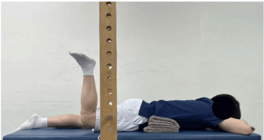
To standardize the hip extension angle, a target bar was used. First, the target bar was positioned next to the participant’s right thigh in the prone position, maintaining a 30-degree abduction of the hip. Then, using a goniometer, the height of the target bar was adjusted to 5 degrees of hip extension. After this, the participants performed hip extension exercises in the prone position, raising their leg until it reached the pre-set height of the target bar.7
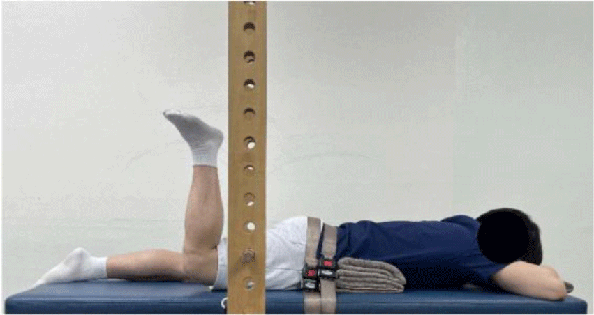
Participants performed PHE under two conditions: (1) no fixation (NONE) (Fig. 1) and (2) fixation on the pelvic (FP) (Fig. 2). For the FP condition, external fixation was applied to the pelvis by positioning a non-elastic belt horizontally across the posterior superior iliac spine (PSIS) and securing it to the exercise table. To prevent compensatory movements, such as excessive lumbar extension and anterior pelvic tilt, which may occur due to instability in the lumbopelvic region, a towel was used. The towel was positioned between the line connecting the xiphoid process of the sternum and both anterior superior iliac spines. The lumbopelvic region of all participants was positioned in a neutral alignment.
First, to standardize the muscle contractions of the GM, both MF and BF, MVIC was measured in the prone position. For the GM, MVIC was measured in the prone position with the knee joint at 90 degrees, while resistance was applied to the distal thigh during active hip extension. For the BF, the measurement was taken in the prone position during active knee flexion, with the examiner stabilizing the thigh and applying resistance to the ankle.20 For both MF, MVIC was measured during active extension of the lumbar spine in the prone position, with resistance applied to the scapula.21 Before the experiment, the order of measurements was randomly assigned using Microsoft Excel (Microsoft, Redmond, WA, USA) to determine the sequence of applying the two different external fixation conditions. Participants were allowed to practice the exercise posture to familiarize themselves with and without the external fixation conditions and the exercises. After a 10-minute practice session, the external fixation condition was set according to the randomized order, and muscle activity for the four muscles was measured during hip extension up to the height of the target bar (Figure 1 and 2). All measurements were performed three times for 5 seconds each, using a metronome set to 60 beats per minute.6
The collected data were statistically processed using the SPSS Version 20.0 software (SPSS Inc., USA). The Shapiro-Wilk test was used to check for normality. A paired t-test was conducted to compare the changes in muscle activity and ratios of the GM, both MF and BF under different external fixation conditions. The level of statistical significance was set at ?=0.05.
RESULTS
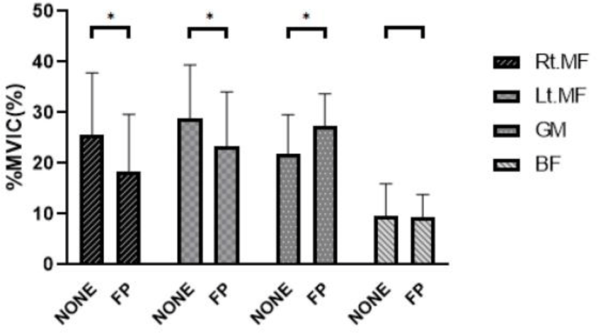
The changes in muscle activity of the GM, both MF and BF during PHE under different external fixation conditions are as follows (Table 2, Figure 3). The muscle activity of the GM significantly increased in the FP condition compared to the NONE condition (Effect size: 0.79, p<0.001). In addition, the muscle activity of both MF significantly decreased in the FP condition compared to the NONE condition (Table 2, Figure 3). However, there were no significant changes in the muscle activity of the BF between the NONE and the FP conditions (p=0.525) (Table 2, Figure 3).

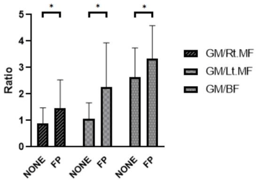
The ratio of muscle activation between the GM and both MF during PHE significantly increased in the FP condition compared to the NONE condition (Table 3, Figure 4). Similarly, the GM/BF activity ratio also during PHE significantly increased in the FP condition compared to the NONE condition (Effect size:0.60, p=0.001) (Table 3, Figure 4).
| Muscle ratio | NONE | FP | t | p | Effect size |
|---|---|---|---|---|---|
| GM/Rt.MF | 0.88±0.59 | 1.48±1.06 | –4.375 | 0.001* | 0.70 |
| GM/Lt.MF | 1.05±0.61 | 2.26±1.67 | –3.274 | 0.006* | 0.96 |
| GM/BF | 2.63±1.10 | 3.34±1.25 | –4.499 | 0.001* | 0.60 |

DISCUSSION
In this study, we aimed to investigate how two different external fixation conditions affect GM muscle activity and the GM/BF activity ratio during PHE. The findings revealed that GM muscle activity increased by 25.23% in the FP condition compared to the NONE condition. Additionally, the GM/BF activity ratio showed a significant increase of 26.99% in the FP condition compared to the NONE condition.
There were various explanations for the result of this study. First, the higher muscle activation of the GM in the FP condition compared to the NONE condition suggests that external fixation contributed to the stability of the lower back and pelvis. This contributed to a more precise hip extension movement, leading to more effective recruitment of the GM.16 It is anticipated that incorporating core stability exercises alongside external fixation could further enhance the functional performance of lower limb movements by facilitating intrinsic stabilization. Similarly, in the Jeon15 study, it was reported that during hip abduction in a standing position, the muscle activity of the gluteus medius significantly increased when external support was provided, compared to when no external fixation was applied. This can be explained by the increased deep stability provided by the external support, allowing the muscles to be activated more efficiently. Furthermore, the Jeon22 study also found that in a group with weak isometric core strength, providing external support resulted in a statistically significant increase in the strength of the hip flexor muscles. This was attributed to the improved core stability from the external support in the lumbopelvic region, enhancing the interaction between the iliopsoas and rectus femoris muscles. Previous studies reported that the application of external pelvic compression in the chronic low back pain group was shown to reduce pain and decrease trunk and hip muscle activity. This suggests that external pelvic compression can help alleviate pain and prevent excessive use of trunk and hip extensor muscles in the chronic low back pain group.14 In contrast, our study found that external fixation of the pelvis during PHE contributed to the activation of the GM, an agonist of hip extension, in healthy subjects. Thus, it suggests that external stabilization during PHE may affect the activation or inhibition of target muscles.
Second, the decrease in multifidus muscle activity in the FP condition compared to the NONE condition in this study is considered to be due to the external fixation applied to the pelvis, which likely controlled compensatory actions such as excessive extension and rotation of the lumbar region caused by synergistic muscles. Although direct comparison is difficult because of different extremities. Hwang and Jeon23 study reported that applying external fixation to the shoulder reduced compensatory actions by the levator scapulae during shoulder flexion. Previous studies explained that the results of this study are due to the external fixation applied to the shoulder, which controlled the compensatory actions of synergistic muscles.
The lack of significant difference in BF muscle activity between the two environments can be attributed to the fact that the PHE was performed in a 90-degree knee flexion position. This position contributes to active insufficiency of the BF, which in turn maximizes the muscle activity of the GM.24
The GM/MF activity ratio increased as the muscle activity of the MF significantly decreased and the muscle activity of the GM significantly increased. Also, despite there being no significant difference in the muscle activity of the BF, the muscle activity of the GM significantly increased, leading to a significant increase in the GM/BF activity ratio. Synergistic muscles work together and influence each other through movement patterns.25 Assuming the movement occurs within the same range of motion, increasing the EMG amplitude of one muscle can improve movement efficiency and reduce the workload of other muscles.26,27 The results of this study suggest that external stabilization applied to the pelvis during PHE not only effectively enhances GM activation but also contributes to pelvic stabilization, helping to reduce overactivation and compensatory actions of adjacent muscles. This finding shows that external pelvic fixation significantly facilitates GM muscle activation and the GM/BF activity ratio during PHE. Therefore, the FP condition during PHE can be suggested as an exercise to specifically activate the GM muscles.
The limitations of this study are as follows. First, the study was conducted exclusively on adult males, limiting the findings’ generalizability. Future studies should include a broader range of participants, including individuals of different age groups, females, and patients with chronic low back pain. Second, the study did not examine the initiation timing and rhythm of GM muscle contractions. Future studies should investigate the relationship between the initiation timing of GM contractions and external fixation. Third, since surface electromyography was used in this study, there is a possibility of crosstalk from adjacent muscles.


