INTRODUCTION
Osteoarthritis (OA) represents a chronic musculoskeletal disorder and a leading cause of worldwide disability.1 OA is a disease caused by damage to joint cartilage and soft tissue and is classified as a representative disease that generally affects the knee, causing pain, functional limitations, and a decrease in quality of life.2,3 Diagnosing OA necessitates that patients present themselves at a medical facility for an evaluation, particularly when indicative symptoms are self-identified through self-diagnosis. However, diagnosing OA early remains challenging due to its dependence on the subjective judgment and feelings of the patient. In addition, most evaluations and therapeutic interventions for degenerative OA are performed after the disease has progressed significantly, so the improvement effect is very low.4 Therefore, it is essential to prevent OA before it occurs and to diagnose it early after it occurs, and there is a need to obtain information about OA without visiting a hospital.
Currently, because there isn’t a clear definition for early OA, it’s hard to diagnose it early and use treatments to slow its progression. Some definitions exist in the literature, and early OA can be described in three ways: early symptoms, early onset in young adults, and initial radiological changes [Kellgren and Lawrence grades 0-1-2].5,6 Additionally, OA has different types, but their definitions are still not consistent.7,8 For this reason, the Early Osteoarthritis Questionnaire (EOAQ) was developed to assess and monitor the clinical progression of early knee OA.9 The EOAQ includes questions about the initial perception of pain or discomfort during daily activities, as well as instances of the knee locking or giving way. For each question, respondents could choose from three options based on the past six months: “not at all,” “rarely (1–3 times),” or “often (more than 3 times)”. Since there are currently no other tools available to assess early OA, despite its limited validation, the adoption of this questionnaire is strongly encouraged to facilitate earlier diagnosis and intervention. It is also necessary to have a guide to determine whether a pharmacological or non-pharmacological treatment is appropriate. Therefore, if we can determine whether EOAQ is associated with the biomechanical properties of early OA, the tool will help clinicians monitor symptoms to prevent disease progression through non-pharmacological treatments or lifestyle changes.
One potential therapeutic focus for delaying the start of knee OA would be to address the biomechanical characteristics of knee OA. The development and progression of knee OA are significantly influenced by varus knee alignment, which is linked to the biomechanical stress exerted on the knee joints.10,11 Varus thrust is thought to be able to significantly raise medial tibiofemoral loading by causing an unexpected lateral shift of the knee.12 The presence of varus thrust is visualized by rapid lateral movement of the knee during the stance, with a return to less varus alignment during functional movement.13 Recent meta-analyses have found that the presence of varus thrust at baseline is associated with almost two-fold greater odds of medial tibiofemoral OA disease progression.14 As varus thrust can occur throughout knee OA, early identification and intervention may have a significant impact on pain and the course of the illness.15,16 Since varus thrust may be a clinical indicator of excessive medial joint load, the significant correlations observed with pain and the progression of OA disease in those with the presence of varus thrust may indicate a relationship between pain and joint load.17
Although several studies have investigated the relationship between the biomechanical characteristics of knee OA and knee OA questionnaires,18-20 there is still no research on early knee OA. Therefore, the purpose of this study was to determine whether EOAQ can reflect the biomechanical characteristics that appear during functional movements in individuals at risk for early knee OA. We divided participants into an early OA risk group and a control group using the EOAQ. We then measured horizontal displacement of the pelvis (PHD), knee (KHD), and ankle (AHD) during functional movements. We hypothesized that the experimental group would exhibit greater PHD, KHD, and AHD compared to the control group.
METHODS
Forty-three manufacturing workers aged 40 to 70 years who are susceptible to knee OA participated in this study, and a total of 86 legs were recruited. Manufacturing workers were chosen because previous studies have shown they face additional risk due to the nature of their work, which often requires them to stand.21 Through the EOAQ, those who answered “frequently” or “rarely” in questions 1 and 2 (questions assessing symptoms of degenerative arthritis) were selected as the experimental group and those who answered “never” were selected as the control group. There were 42 legs (24 males, 18 females) in the experimental group and 44 legs (22 males, 22 females) in the control group. Table 1 summarizes the characteristics of the participants in each group. Subjects were excluded if they had experienced a lower extremity injury in the past 6 months or had previously been diagnosed with hip surgery, rheumatoid arthritis, or neurological conditions. All subjects were informed about the procedures of this study and provided an informed consent form before the experiment. This study was approved by the Sangji University Institutional Review Board.
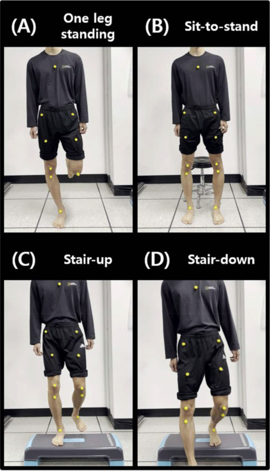
A regular smartphone (iPhone 15; Apple Inc., USA) with video recording capabilities (4K, 2,556 × 1,179 pixels at 240 fps) was used to assess PHD, KHD, and AHD during one-leg-standing (OLS), sit-to-stand (STS), stair-up (SU), and stair-down (SD) in the frontal plane. The attachment locations for reflective markers were as follows: the anterior superior iliac spine for the pelvis, the middle of the patella for the knee, and the top of the navicular bones for the ankle. The camera was positioned on a tripod that was 60 cm in height and placed 250 cm in front of the participants. A 15-cm high step box was used for SU and SD.
The PHD, KHD, and AHD during OLS, STS, SU, and SD were analyzed using Kinovea software. All recorded videos were analyzed with the stable version of Kinovea (v. 0.8.16, Kinovea, Bordeaux, France). Kinovea is a free 2D motion analysis software that enables the establishment of kinematics parameters. In this study, the horizontal displacement at the highest outward deviation was recorded. PHD was excluded because the marker is obscured when sitting.
The participants were asked to perform two trials (right and left leg), three times of the OLS, STS, SU, and SD test. Since the EOAQ includes questions about pain or discomfort during daily activities, functional tests related to these activities were used for evaluation. Before the records, the participants were familiarized with the testing protocol, provided instructions, and asked to practice the tests to ensure proper motion. Each participant took off their shoes to avoid variability in different sole materials. The order of the tests and tested leg was random. Subjects were given sufficient rest between trials to avoid fatigue. For the OLS test, subjects were instructed to stand quietly in the upright position with the foot parallel at hip width. The subject stands upright for 10 seconds and the other knee flexed 90° (Figure 1A).22 For the STS test, participants were instructed to sit with the foot and knee parallel at hip width. The participants rose to stand at their preferred speed for three trials while watching a standing eye-level target. The participants chose the initial foot position, which was maintained throughout. Rising to stand commenced with arms by the side, however, arms were free to move during rising (Figure 1B).23 For the SU test, participants were instructed to place one leg on a step box with keep the hip, knee, and foot parallel. And, participants were provided with both a demonstration and verbal instruction on performing the step-up without specific directions on knee and hip alignment (Figure 1C). The SU test was completed by raising the non-positioned leg until the participant’s heel lightly touched the floor of the 20 cm height step box. For the SD test, participants were instructed to sit with the foot and knee parallel at hip width. Participants were provided both a demonstration and verbal instruction on performing the step down without specific directions on knee and hip alignment. The SD test was completed by lowering their non-stance leg until the participant’s heel lightly touched the floor in front of the 20 cm height step box (Figure 1D).24 During four functional tests, horizontal displacement of the lower extremities were video-recorded and analyzed using the Kinovea. The PHD could not be measured in the STS test because the markers attached to the pelvis were obscured in the seated position.
Statistical analyses were performed using the SPSS for Windows (ver. 25.0; IBM Co., Armonk, NY, USA). All data were tested for normal distribution using the Kolmogorov-Smirnov test. An independent t-test was conducted to identify significant differences in the PHD, KHD, and AHD during OLS, STS, SU, and SD between the experimental and control groups. The level of significance was set at p<0.05.
RESULTS
The results of the independent t-test indicated that the legs of the experimental group had statistically greater horizontal displacement in all variables (p<0.001) during the OLS test compared to the control group. In the STS test, the legs of the experimental group had statistically greater KHD (p<0.001). However, there were no significant differences in AHD during the STS test. The legs of the experimental group had statistically greater PHD (p=0.047), KHD (p<0.001), and AHD (p<0.001) during the SU test compared to the control group (Table 2). In the SD test, the legs of the experimental group statistically greater PHD (p=0.003), KHD (p=0.031), and AHD (p<0.001) compared to the control group (Table 2).
DISCUSSION
The purpose of this study was to determine whether the EOAQ can reflect the biomechanical characteristics of knee OA. The experimental group exhibited greater lateral horizontal displacement in most variables than the control group. Therefore, considering the results of this study, it can be suggested that the EOAQ reflects the biomechanical characteristics of knee OA, particularly concerning varus thrust. Additionally, although verification of the use of the EOAQ for early diagnosis and prevention of knee OA was limited,9 the results of this experiment can serve as evidence to some extent.
There have already been many studies investigating the biomechanical characteristics of knee OA during functional tests, and attempts have also been made to correlate these characteristics with questionnaire responses.18 Most studies have shown that increased horizontal displacement, such as hip lateral sway and knee varus thrust, that occurs during functional movement is related to knee OA.14,25,26 However, previous studies focused on patients who already had OA, so they do not fully explain the biomechanical characteristics of early knee OA or how it should be prevented. This study is the first attempt to address the aforementioned limitations and investigate whether the EOAQ can reflect the biomechanical characteristics of knee OA. The results of our study showed that participants at risk of early knee OA exhibited greater varus thrust in the hip and knee, consistent with previous studies investigating the biomechanical characteristics of knee OA. Therefore, the results of these studies emphasize the importance of considering varus thrust in understanding and managing early knee OA. By identifying specific movement characteristics early, it is possible to potentially influence disease progression and improve long-term outcomes for individuals at risk of developing knee OA.
Among the variables, only AHD during the STS showed no significant difference between groups. The findings were inconsistent with a previous study that reported higher ankle varus moments in the OA group.27 Several possibilities may explain these results. While those studies focused on OA patients, our research targeted individuals at risk of knee OA using the EOAQ. Previous studies that compared the biomechanical changes between the asymptomatic group and the moderate and severe OA groups reported that kinematic differences in the ankle joint were only observed in the severe OA group.28 Therefore, differences may not be evident between the groups in our study. Another potential explanation is the alteration in foot arch dynamics observed during the STS test. Considering that the initial position of the STS test is a seated posture that does not bear weight, the transition to a standing posture may exert a greater influence on the modifications of the foot arch. The functional navicular drop test also measures the differences between weight-bearing and non-weight-bearing conditions in similar contexts,29 which may render the measurements of ankle movement during the STS test less meaningful.
Our study had several limitations. First, only the horizontal displacement of the lower extremities was measured. Future research should include measurements of rotational movements that occur during functional movement tests. Second, this study was a cross-sectional study. Future research is needed to determine whether EOAQ responses change after interventions aimed at preventing lateral horizontal displacement. Third, an unexpectedly high number of participants were recruited. While a larger sample size can enhance the robustness of the findings, it also introduces several challenges related to potential data bias. Another limitation is the difficulty in generalizing the results. The participants were all manufacturing workers aged between 40 and 70. Further research is needed to determine if similar results can be observed in different occupational groups and age ranges.
CONCLUSIONS
The results of this study indicate that the EOAQ reflects lateral horizontal displacement, a biomechanical characteristic of knee OA. The observed differences in varus thrust between groups during the OLS, STS, SU, and SD tests suggest that varus thrust should be considered in the early management and prevention of knee OA. Additionally, this study presents the need and potential for integrating the EOAQ with other diagnostic tools to improve the detection of early knee OA.


