INTRODUCTION
One of the causes of scapular dyskinesis, scapular downward rotation syndrome (SDRS), is when the scapular medial border is not parallel to the spine, the inferior angle of the scapula is located more inside than the superior angle of that, and the distance between the scapular medial border and the spine is narrowed to 7 to 8 centimeters or less.1,2 This is also considered to be one of the classifications of impaired scapular alignment.1 Scapular dyskinesis is observed in patients with rotator cuff injury, shoulder instability, and shoulder impingement syndrome, and has been reported to be associated with labral injury and acromioclavicular joint injury.3–5
SDRS is characterized by shortening of the muscles that downward rotate the scapula, such as levator scapula, rhomboid, latissimus dorsi, and pectoralis major (PM), and weakness of the muscles that upward rotate the scapula, such as the upper trapezius, serratus anterior (SA), and lower trapezius.6–8 Especially, a notable common feature of subjects with SDRS is insufficient scapular upward rotation due to serratus muscle weakness.9 Insufficient scapular upward rotation during flexion of the shoulder reduces the activation of the SA and leads to overactivity of the excessive PM as a compensatory effect for the weakening of the SA.4,10 The PM contracts isometrically when the shoulder joint is moved, stabilizing the glenohumeral joint, clavicle, and scapula. However, the underactivity of the SA leads to the overactivity of the PM.11 For this reason, previous studies have shown that SDRS is responsible for a variety of injuries such as movement impairment, shoulder impingement syndrome, tendinitis and rupture of the supraspinatus or rotator cuff, thoracic outlet syndrome and nerve entrap-ment, subluxation and instability of the humerus, and pain in the acromioclavicular joint or sternoclavicular joint.1,2
A major characteristic of subjects with SDRS is the insufficient upward rotation of the scapula due to the weakness of the SA. A popular exercise clinically used to effectively strengthen the SA in many studies is the Push-Up Plus exercise.12–15 The serratus punch exercise is also used a lot.10,16 When comparing the muscle activities of the SA and PM during the serratus punch exercise performed in a standing position, the modified push-up plus wall version and the modified knee push-up plus floor version, the serratus punch exercise were found to have higher muscle activities of the SA.10
Previous studies on SDRS have primarily sought to determine changes in pain and muscle activity between the control and experimental groups. However, there is a lack of studies that suggest the best exercise position for SDRS by comparing the SA and the PM according to the positions for SA strengthening exercise. Therefore, this study is intended to determine the most effective exercise position for SDRS by comparing the muscle activities around the shoulder according to the positions for SA strengthening exercises. We hypothesized that there would be a difference in the ratio of muscle activity between the SA and the PM according to the SA strengthening exercise position for SDRS, and we expected that the position that increases the muscle activity of the SA more than the PM was the side-lying position.
METHODS
Thirty-nine individuals with SDRS participated in the study (Table 1). Inclusion criteria were (1) the inferior angle of the scapula is located inside the superior angle of the scapula (2) the acromioclavicular joint is lower than the sternoclavicular joint and (3) an individual whose joint range of motion is less than the normal range when flexion the shoulder joint, so that the inferior angle of the scapula doesn’t reach the midaxillary line. Exclusion criteria were (1) medical history of shoulder surgery and (2) pain in the shoulder joint.2 This study was approved by the institutional review board of Kyungsung University (KSU-21-02-003).
| Variable | Subject (n=39) |
|---|---|
| Age (years) | 29.70±4.02 |
| Height (cm) | 167.70±7.82 |
| Weight (kg) | 63.01±11.54 |
| Gender (male/female) | 20/19 |
| Amounts of SDRI (cm) | 0.83±2.42 |
For normalization of the muscle activity of each muscle, maximal voluntary isometric contraction (MVIC) was measured, and surface electromyography (EMG) (Noraxon TeleMyo DTS wireless system, Noraxon, USA) was used, the notch filter was set to 60 Hz. The band pass filter was set at 10–450 Hz, and the sampling rate at 1,000 Hz. In this study, muscle activities of the SA and PM were measured. EMG patches were applied by following the guidelines of Criswell et al.17–19 Before placing the elec-trodes, the area where electrodes are to be attached was shaved. Then, the skin was cleaned with an alcohol swab. Two separated bipolar surface electrodes were placed 2 cm apart. SA was attached midaxillary line at the same height as the scapular inferior angle, PM was attached 2 cm inside the axillary fold. Muscle tests for MVIC of SA were performed following the guideline of Bayattork et al.20 Muscle tests for the MVIC of PM were performed according to the guideline of Lehman et al.21 Muscle contraction was standardized using %MVIC. The signal of muscle activities was recorded while each participant was maintaining scapular strengthening exercises in three different positions for 5 seconds. The signals at 3 seconds were analyzed, excluding each 1 second at the beginning and end of the exercise. The average EMG data was collected, the average root mean square (RMS) value was calculated, and the average of the three trials was used for the anlaysis.
All subjects in this study performed scapular strengthening exercises in three different positions to compare muscle activities around the shoulder according to the exercise positions, and the subjects’ exercise positions were executed after random assignment.
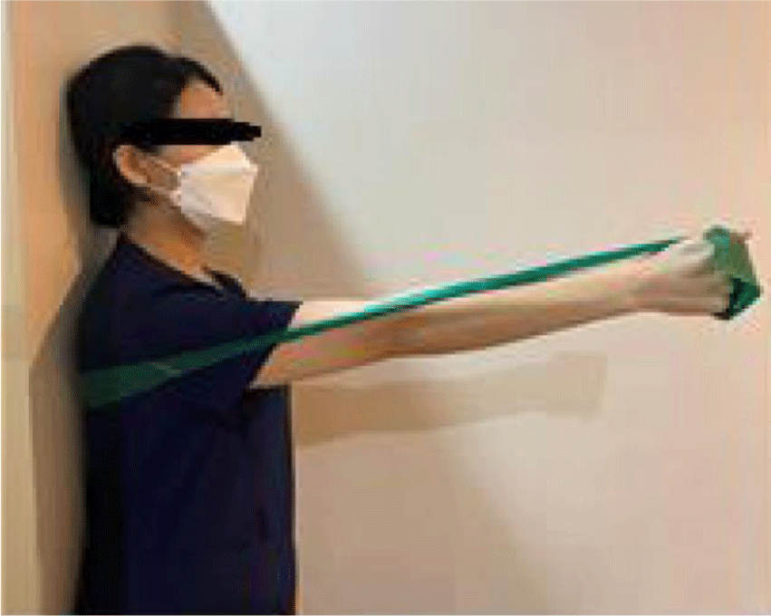
In an upright position, subjects were secured against a wall with a green elastic band (Thera-band) wrapped around the most protruding part of the back when rounding their back, as determined by the principal investigator, on the side with the scapular downward rotation, and the shoulder flexion was at a 90° angle.22,23 The humeral elevation angle was measured using a goniometer, and after the target point was placed on the plumb line, scapular protraction was performed toward the target point during the exercise.16 Participants were instructed not to turn their body to one side and to protract their scapula as far as possible (Figure 1).24 The tension of the elastic band was determined by each participant's ability to perform at least 10 repetitions in the set position.25,26
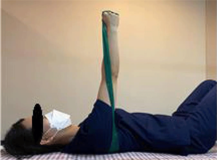
In a supine position, the subjects fixed the elastic band to the floor using their body weight at the same location as in the condition of the scapular strengthening exercise in a standing position. Then, they performed shoulder flexion at a 90° angle while holding the elastic band.22,23 They performed the same exercises for scapular strengthening in the standing position (Figure 2).
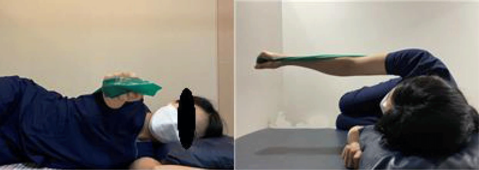
In a side-lying position with the shoulder with scapular downward rotation toward the ceiling, the subjects fixed the elastic band to the floor using their body weight at the same location as in the condition of scapular strengthening exercise in a standing position. To maintain a consistent lateral trunk position, the external auditory meatus was visually aligned with the midline of the lateral thorax and the greater trochanter of the hip, by the ideal alignment proposed by Magee.27 Then, they performed shoulder flexion at a 90° angle while holding the elastic band.22,23 The experimenter monitored the trunk position throughout the exercise to prevent any flexion or rotation of the trunk in the subjects.28 They performed the same exercises for scapular strengthening exercise in the standing position (Figure 3).
This research used SPSS ver 26.0 for data analysis. Repeated measures one-way analysis of variance was used to compare muscle activities of the SA and PM muscles during shoulder-strengthening exercises. If significant interactions were confirmed among the exercises, the Bonferroni correction was set at 0.017 (0.05/3) for statistical significance.
RESULTS
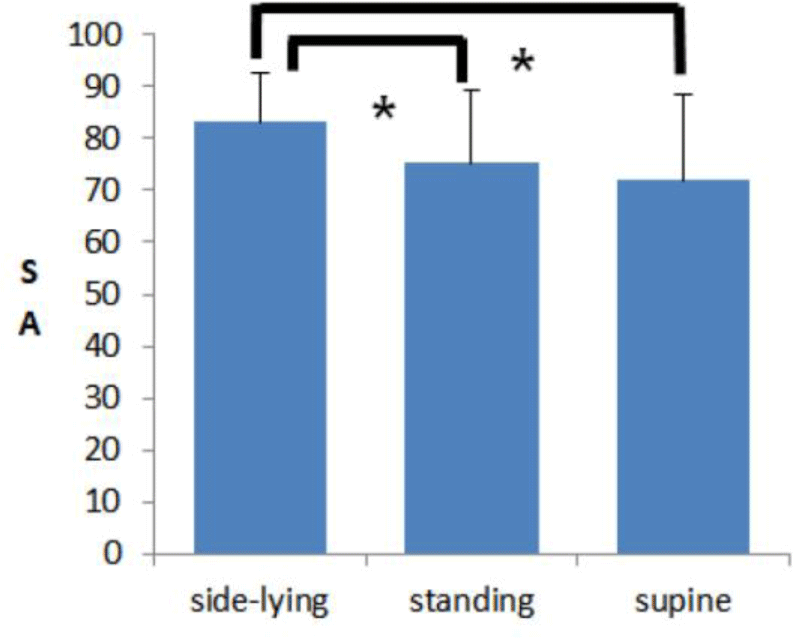
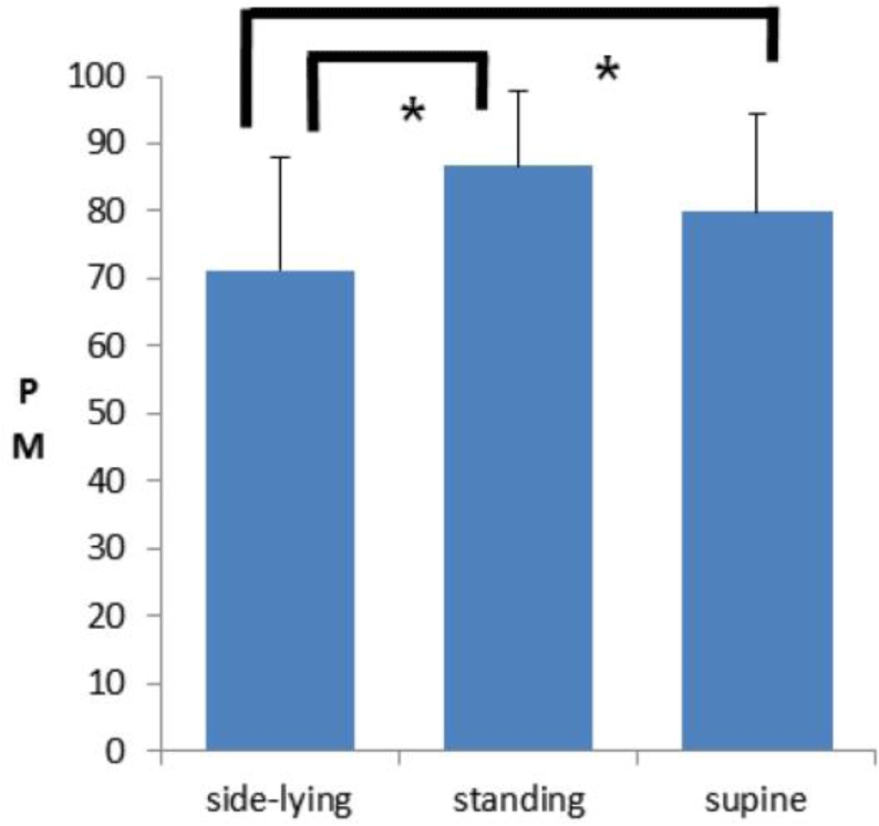
When the muscle activities of the SA were compared by position during shoulder-strengthening exercise, the muscle activity of the SA increased in the side-lying position compared with the standing and supine positions, showing a statistically significant difference (p<0.05) (Table 2). When the muscle activities of the PM were compared by position during shoulder-strengthening exercise, the muscle activity of the PM decreased in the side-lying position compared with the standing and supine positions; there was a statistically significant difference (p<0.05) (Table 2).
| Muscle | Mean±SD (%MVIC) | F value | p value | ||
|---|---|---|---|---|---|
| Side-lying | Standing | Supine | |||
| SA | 83.17±9.23 | 75.25±14.02 | 71.95±16.52 | 6.59 | 0.002* |
| PM | 71.20±16.65 | 86.69±11.11 | 79.85±14.40 | 9.19 | 0.001* |
When comparing the muscle activity ratio of the SA to the PM by position during scapular strengthening exercises, the muscle activity ratio of the SA increased in the side-lying position compared with that of the standing and supine positions; the difference was statistically significant (p<0.05) (Table 3).
| Muscle | Positions | F value | p value | ||
|---|---|---|---|---|---|
| Side-lying | Standing | Supine | |||
| SA/PM | 1.17 | 0.87 | 0.90 | 13.05 | 0.00* |
In the Bonferroni post-hoc analysis, SA muscle activity in the side-lying position exhibited statistically significant differences between standing (p<0.014) and supine position (p<0.004) (Figure 4). PM muscle activity in the side-lying position exhibited statistically significant differences between standing (p<0.001) and supine position (p<0.012) (Figure 5).
DISCUSSION
This study analyzed the muscle activities around the shoulder during elastic band-applied scapular strengthening exercises in 39 adults with SDRS by exercise position to determine the best exercise position to strengthen the SA appropriate for SDRS intensively. Elastic band-applied Scapular strengthening exercises were performed in the side-lying, standing, and supine positions.22
The results showed that the side-lying position signifi-cantly increased the muscle activity of the SA compared with that of the standing and supine positions during scapular strengthening exercises for SDRS, and the standing position increased the muscle activity of the SA compared with that of the supine position, but there was no significant difference.
The reason the muscle activity of the SA is lower during scapular strengthening exercise in the supine position than in other positions. Ekstrom et al.29 reported that during a test where participants applied resistance with their hands and elbows while scapular protraction in a supine position with the shoulder flexed to 90 degrees, the SA EMG activity was only 57% of the MVIC. The low muscle activation of the SA during scapular protraction in the supine position was attributed to the scapula being fixed in place, which leads to co-contraction of the surrounding scapular muscles.29 Scapular protraction occurs due to the contraction of the PM, which pulls the scapula and humerus, counteracting the action of the SA.30 Based on the results of this study, it is deemed that scapular protraction in a supine position may cause co-contraction of the PM and the SA muscle, limiting the function of the SA and the scapular movement. It is shown that even though the scapula is protracted to its maximum range in the supine position, it remains in place, indicating co-contraction of the muscles around the scapula.20
Providing resistance during active scapular protraction in the side-lying position increases the muscle activity of the SA, which may help treat scapular dysfunction.25 Muscle activity can be defined as a total of four levels: low (<20 %MVIC), moderate (20–40 %MVIC), high (41–60 %MVIC), and very high (>60 %MVIC).31 For normal individuals, the muscle activity for the SA during wall press exercise with bare hands in the standing position and bench press exercise with bare hands in the supine position was measured to be 23.3%±13.4% MVIC for the wall press exercise and 36.6%±14.3% MVIC for the bench press exercise, when performed at 80% of the MVIC.32 This indicates that performing exercises without resistance is insufficient for strengthening the SA, and appropriate resistance is necessary for effective SA strengthening.28 According to Hintermeister et al.32 seven exercises—internal rotation, external rotation, seated rowing, narrow grip, middle grip, wide grip, forward punch, and shoulder shrug—were performed to compare the activity of seven muscles using elastic bands. The results showed that the forward punch exercise elicited the greatest muscle activity in five muscles: the serratus anterior, pectoralis major, infraspinatus, supraspinatus, and anterior deltoid. These findings suggest that shoulder exercises using elastic band resistance are effective.33 In this study, the muscle activity of the SA was 83.17%±9.23% MVIC during scapular strengthening exercises in the side-lying position using an elastic band. From the results of this study, the higher SA activation observed in the side-lying position, compared to the standing position, is likely due to the ability to selectively strengthen the SA using elastic bands in this posture. Tabott et al.28 reported that scapular protraction exercises in the side-lying position minimize the co-contraction of the surrounding scapular muscles. Scapular protraction exercises in the side-lying position are clinically effective in promoting the activation of the SA, minimizing excessive stress on the shoulder muscles, rotator cuff, and glenohumeral joint while reducing the activity of surrounding scapular stabilizers.28 It is likely that the higher muscle activity in the side-lying position compared to the other two positions in this study is because of the selective strengthening of the SA using elastic bands. Therefore, shoulder-strengthening exercise using elastic band in a side lying position can selectively strengthen the SA for SDRS.
In this study, the side-lying position significantly in-creased the SA/PM ratio during scapular strengthening exercises for SDRS compared with the standing and supine positions. Park et al.26 compared the muscle activity of the shoulder muscles in individuals with scapular winging, a condition characterized by weakness of the SA, during scapular protraction with resistance of scapular abduction using an elastic band in a standing position and during scapular protraction without resistance. As a result, it was reported that scapular protraction with resistance of scapular abduction increased the muscle activity of the SA and decreased the muscle activity of the PM.33 Therefore, scapular strengthening exercises using elastic bands can selectively activate the muscle activity of the SA in subjects with SDRS because they can perform the exercise according to individual scapular exercise abilities.
There are a few limitations to this study. First, the age range is limited to young adults, and it is difficult to generalize the results due to the small number of subjects. Second, muscle activity around the shoulder was measured only at 90° of shoulder flexion. It is necessary to perform scapular strengthening exercises at different angles to compare the effects of these exercises on muscle activity around the shoulder at each angle. Finally, the resistance of the elastic band increases as it is stretched, which makes it impossible to provide consistent resistance values to all participants. Future studies would need to include a larger sample of subjects across different age groups with SDRS and ensure consistent resistance values at various angles during scapular strengthening exercises to compare muscle activity around the shoulder.
CONCLUSIONS
Scapular strengthening exercises in the side-lying position significantly increased the muscle activity of the SA and significantly decreased the muscle activity of the PM compared with the exercises in the other two positions. Therefore, the most effective position to strengthen the SA for SDRS is the side-lying position. This information can be used by clinicians treating SDRS when selective activation of SA is required.


