


INTRODUCTION
The human knee is a complex joint that plays a crucial role in our daily activities, providing stability and flexibility for movements such as walking, running, and jumping.1 Knee alignment refers to the way the bones in the leg are positioned in relation to each other.2 The ideal alignment involves a straight line from the hip, through the knee, to the ankle.3
Deviations from this optimal alignment can occur, with one common misalignment being varus or valgus alignment.4 Varus or valgus knee alignment plays a crucial role in the development and progression of osteoarthritis (OA).5 The knee varus type is relatively more common in OA patients. Knee varus, commonly known as bowlegs, and OA are interconnected through the biomechanical stress placed on the knee joints. Knee varus is a deformity where the knees are positioned outward, causing increased pressure on the inner side of the knee joint.6 This altered alignment can contribute to the development and progression of OA in the affected joints. Over time, the loss of cartilage can contribute to OA, a degenerative joint condition characterized by pain, stiffness, and reduced joint function.7
OA, a degenerative joint condition, affects millions of people worldwide, impacting their quality of life and mobility.8 While it is a prevalent condition, the importance of early detection in OA cannot be overstated. Early identification allows for timely intervention, which can significantly influence the progression of the disease and improve long-term outcomes for individuals.9
Therefore, it is important to understand the movement characteristics of the knee varus type and the stages through which it develops into OA. In particular, it is necessary to understand the characteristics of the varus knee type that causes OA. According to Sahrmann’s diagnostic classification,10 it can be classified into 2 types of knee varus alignment (Type 1: hip internal rotation, knee hyper-extension, tibia internal rotation, Type 2: hip external rotation, slightly knee flexion, tibia external rotation). Type 1 and 2 have similar knee varus alignment, but the mechanism by which varus alignment is formed is different. This difference in alignment formation results in different movement characteristics when performing various functional movements.
However, there is no study comparing the characteristics of movement patterns according to the knee varus alignment type. The purpose of this study is to investigate the characteristics of lower extremity movements during functional activities according to knee varus types. It was hypothesized that there will be different movement characteristics between the two knee varus types.
METHODS
Thirty subjects with knee varus participated in this study (age: 42±3.5 years, height: 171±10.1 cm, weight: 69.3±8.9 kg). Before the participation of this study, subjects were classified into two knee varus type.10 It can be classified into two types of knee varus alignment (Type 1: hip internal rotation, knee hyper-extension, tibia internal rotation, Type 2: hip external rotation, slightly knee flexion, tibia external rotation) (Figure 1A). Fifteen subjects were assigned to each group. Subjects are excluded if they have experienced a lower extremity injury in the past 6 months or have previously been diagnosed with hip surgery, rheumatoid arthritis, osteoarthritis, or neurological conditions. All subjects were told about the procedures of this study and offered informed consent form before the experiment. This study was approved by the Sangji University Institutional Review Board.
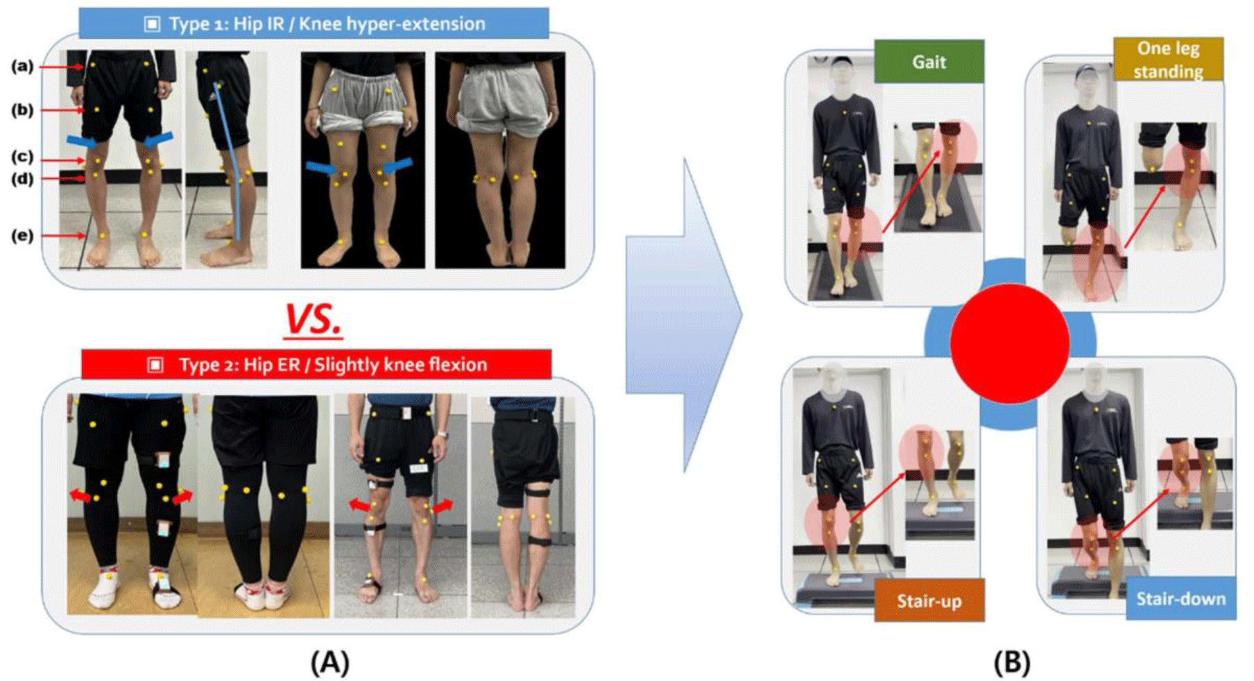
The movements of the subjects’ lower extremity (hip and knee) during 4 functional (gait, one leg standing, stair-up, and stair-down) tests were recorded using a smartphone (Iphone 15, Apple corp., U.S.A). An adjustable tripod was placed 1 m in front of the participants and placed at the level of the knee joint. Round yellow markers of 1 cm in diameter were attached on the anterior superior iliac spine (ASIS), mid-point of femur, mid-point of patellar, tibial tuberosity, and dorsum of ankle (Figure 1).
Gait was performed on a walking pad. The speed of the walking pad was set at 1.5 m/s considering the subjects’ risk of falling, and data were collected for 5 seconds. One leg standing test was conducted on firm ground. A participant stands on one leg with the contralateral knee flexion 90° for 5 s. Both hands were positioned parallel to the body. The participants were kept as balanced as possible until there was a signal to stop with the signal to start the experimenter. The experiment was stopped when an unbalanced posture (unsupported feet touched the ground or the torso was tilted excessively) from the starting position. The stair up test started with one foot placed on a 20 cm height box. Subjects were instructed to climb the stairs at a comfortable pace according to the researcher’s instructions. The stair down test started with both feet placed on a 20 cm height box. According to the researcher’s instructions, the foot to be measured is placed on the box and the other foot is lowered to the ground. Subjects were instructed to descend at a comfortable speed. During 4 functional tests, lower extremity movements (vertical/horizontal displacement of ASIS, midpoint of the patellar, dorsum of ankle) were video-recorded and then analyzed using Kinovea software (Figure 1B).11 The maximum vertical/horizontal movement distance that the marker moved from the beginning to the end of each test was calculated. Kinovea© is a free 2D motion analysis software that enables the establishment of kinematics parameters. All recorded videos were analyzed with the Kinovea (v. 0.8.15, Kinovea, Bordeaux, France).
Statistical analyses were performed using the SPSS for Windows (ver. 25.0 software; IBM Co., Armonk, Ny, USA). To verify the normality of data distribution, Shapiro-Wilk test wad used. An independent t-test was used to compare the characteristics (vertical/horizontal displacement of hip/ knee joints) between type 1 and 2. The level of statistical significance was set at α of 0.05.
RESULTS
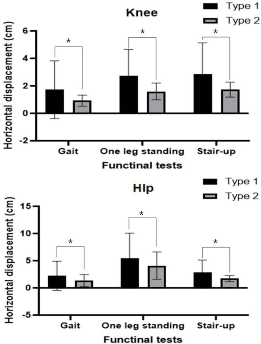
The Shapiro-Wilk test presented the normality of the data (p>0.05). There were significant differences in lower extremity kinematics between the two types. Knee varus thrust (horizontal displacement), hip lateral sway (horizontal displacement), and movement were significantly increased in type 1 in gait, one-leg standing, and stair up tests (Figure 2). Knee varus thrust is defined as the outward bending of the knee that occurs during walking and various functional movements, and hip lateral sway also refers to the phenomenon in which the hip joint moves outward from the center of the body during various functional movements.12, 13
DISCUSSION
Understanding alignment and movement patterns in subjects with OA is of importance in advancing knowledge of this exacerbate condition and refining therapeutic approaches.12 The human body’s alignment, particularly in weight-bearing joints like the knees, significantly influences joint mechanics and the distribution of forces during movement.13 In individuals with OA, deviations from optimal alignment can exacerbate biomechanical stress, contributing to the degeneration of joint structures.14 Investigating specific movement patterns associated with OA provides critical insights into the progression of the disease and helps identify modifiable factors that may influence its course.15 The people with knee varus showed greater knee external adduction moments, knee adduction, eversion, and lateral ground reaction force than the people with normal knee alignment during walking. Additionally, those with knee varus presented increased knee flexion and external knee flexor moments during midstance phase.16 These biomechanical changes cause structural changes in the knee joint, resulting in medial knee OA.17
The purpose of this study was to investigate the characteristics of lower extremity movements during functional activities according to knee varus types. It was found that there was a significant difference in lower extremity kinematics between the two types. Knee varus thrust (horizontal displacement), hip lateral sway (horizontal displacement), and movement were significantly increased in type 1 in gait, one-leg standing, and stair up tests. The observed differences in movement characteristics between Type 1 and 2 knee varus alignments can be attributed to the distinct biomechanical mechanisms underlying the formation of these varus types. While both types exhibit a varus alignment, the specific alterations in hip, knee, and tibia positioning differ. For Type 1, the varus alignment is associated with hip internal rotation, knee hyperextension, and tibia internal rotation. In contrast, Type 2 is characterized by hip external rotation, slight knee flexion, and tibia external rotation. These differences in alignment mechanisms lead to variations in how forces are distributed and absorbed during functional movements. For instance, during gait, the increased knee varus thrust observed in Type 1 suggests greater pressure on the inner side of the knee joint due to the outward positioning of the knees. The hip lateral sway, indicative of side-to-side movement, is likely influenced by the internal rotation of the hip in Type 1. These alterations in joint positioning and movement patterns contribute to the observed differences in lower extremity kinematics between the two types. The unique biomechanical characteristics of each knee varus alignment type result in distinct movement patterns during functional activities.10 Understanding these differences is crucial for tailored therapeutic interventions. The findings highlight the importance of identifying specific problems related to abnormal horizontal displacement in Type 1 individuals, emphasizing the need for targeted assessments and interventions to address the challenges associated with this particular varus alignment. Overall, recognizing the nuanced biomechanics of Type 1 and 2 knee varus alignments provides valuable insights for clinicians and researchers working towards developing effective strategies for managing and treating degenerative arthritis in these patients. The findings emphasize the importance of considering knee varus alignment subtypes in understanding and managing OA. Early identification of specific movement characteristics related to knee varus alignment types could inform targeted interventions, potentially influencing disease progression and improving long-term outcomes for individuals at risk of developing OA.
Our study had some limitations. First, only the vertical-horizontal displacement of the foot/knee was measured. Additionally, the change in the activity of lower extremity muscle activity during 4 functional tests was not measured. Future research requires measurement of rotational movements that occur during functional movements and measurement of lower extremities muscle activity. Second, the study was conducted cross-sectionally. further research is warranted to investigation the long-term effects of these distinct movement patterns and their correlation with OA development. Additionally, investigating interventions tailored to each knee varus alignment type may offer new methods for personalized treatments, ultimately enhancing the effectiveness of prevention and therapeutic approaches in managing knee OA.
CONCLUSIONS
This study contributes valuable insights into the movement characteristics associated with different knee varus alignment types, shedding light on potential connections to OA. The observed differences in knee varus thrust, hip lateral sway, and movement between Type 1 and 2 varus alignments during gait, one-leg standing, and stair-up tests suggest distinct biomechanical patterns associated with each alignment type.