


INTRODUCTION
A straight neck is characterized by the loss of the normal lordotic curve of the cervical spine and change into straight or kyphosis.1 Reduced cervical lordosis changes the dynamic kinematics of the cervical spine during cervical flexion and extension.2 This change exerts stress on the cervical spine, which might cause cervical kyphosis by facilitating degenerative changes in the joints.3 Moreover, a straight neck contributes to musculoskeletal pain in the shoulders and neck due to muscle imbalance and ligament tension around the neck.4,5
Several researchers have studied to decrease pain and to correct alignment of the straight neck. Alpayci and Ilter reported that isometric cervical extension exercise for 3 months decreased neck pain and improved neck curvature in patients who had cervical pain with reduced cervical lordosis.6 Similarly, Lee et al. reported that cervical and shoulder retraction exercises decreased neck pain and improved cervical lordosis among patients who had cervical pain with reduced cervical lordosis.7 However, they failed to clearly discuss the mechanism by which these exercises improved cervical lordosis. In addition, the diagnosis and prescriptions provided by physicians for patients who had cervical pain with a straight neck were not designed as a physical therapy program from which evidence could be obtained.
Sahrmann classified cervical patients into those with cervical extension, flexion, rotation, flexion-rotation, and extension-rotation syndrome based on the Movement System Impairment (MSI) of the cervical spine.8 In particular, the classification of the cervical spine focused on confirming abnormal alignment and movement direction causing cervical pain and functional change.8 In line with this, patients with cervical flexion syndrome have been characterized by a straight neck and reduced kyphosis of the thoracic spine.8 Reduced thoracic kyphosis can result in excessive flexion in the relatively more flexible lower cervical spine during cervical flexion, while increasing compression and shear forces in the cervical spine.8 Unfortunately, no studies to date have applied the MSI approach to patients with cervical flexion syndrome with a straight neck so far.
We hypothesize that correction of the patient’s abnormal alignment and movement pattern will decrease the pain and remove the cause of the tissue irritation. Thus, the present study aimed to describe the treatment and prognosis of a patient with neck pain who had been confirmed to have cervical flexion syndrome with a straight neck based on the MSI approach.
CASE HISTORY
Our patient was a 37-year-old man suffering from neck pain for 3 months. He had experienced a tingling sensation in both arms, especially in the 4th and 5th fingers, which prompted his visit to Hansol Hospital in Daegu. He was diagnosed with cervical disc herniation and prescribed physical therapy by a physician. The patient worked as a hair designer for 10 years and reported that this work required repeated and continuous bending of his neck. He had a depressed scapular posture and felt pain on his posterior lower cervical vertebra during cervical flexion, which he rated at 6 out of 10 on the Visual Analogue Scale (VAS) (Table 1).9 In addition, the patient experienced a sharp posterior lower cervical pain during cervical extension. The Neck Disability Index (NDI) was used to assess the cervical functional disability of the patient. The NDI consists of 10 categories, such as pain intensity, headache, personal care, and sleeping and is useful for the assessment of functional limitation level in the daily life of the patient with cervical pain.10 It has appropriate reliability and validity and has scores ranging from 0 to 50 with higher scores indicating greater functional impairment. Before treatment, our patient had an NDI score of 34, which indicated severe disability. This study was approved by the Institutional Review Board in Daegu Health College (DHCIRB-2023-09-002).
5/5, normal strength; 4+/5, able to hold against moderate to strong resistance; 4/5, able to hold against moderate resistance; 4-/5, able to hold against slight to moderate resistance; 3+/5, able to hold against minimal resistance; 3/5, able to hold against gravity but not against additional minimal resistance applied manually; 2+/5, moves through partial range of motion against gravity.
PHYSICAL EXAMINATION
Physical examination was conducted to confirm the patient’s movement impairment according to Sahrmann.8 The first author who is a physical therapist with 19 years of experience conducted the examination. The examiner performed the test in various positions according to the MSI approach and reevaluated the movement impairment after correcting the alignment and movement dysfunctions when symptoms occurred.
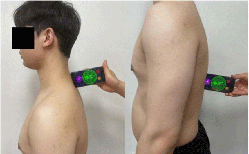
The cervical active range of motion (ROM) was measured using a smartphone (iPhone SE2) with a built-in inclinometer. Smartphone has the cost-effective advantage and can be used easily and conveniently in clinical examinations.11 Goniometer-Pro (5fuf5 CO, Stephanskirchen, Germany) one of the smartphone inclinometer applications has high reliability with an intraclass correlation coefficient of 0.99.12 To measure the cervical active ROM of the patient, he was instructed to sit on a chair with both feet on the floor. The smartphone running Goniometer-Pro was placed right next to the patient’s external auditory meatus. Then, the axis of the app was aligned and the patient’s cervical flexion-extension ROM was measured. To measure cervical lateral flexion ROM, the center of Goniometer-Pro was placed on the C7 spinous process, and one axis of the application was aligned with the occipital protuberance. Finally, to measure cervical rotation ROM, the center of Goniometer-Pro was placed at the center of the patient’s head, after which one axis of the application was aligned with the nose.13 Each ROM measurement was performed a total of three times and, the average values were used (Table 1).
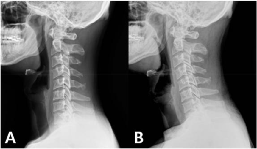
The alignment of the thoracic spine was also measured in the standing position using the smartphone. The examiner placed the midpoint of the bottom of the smartphone running Goniometer-Pro on the T1 spinous process and T12 spinous process of the thoracic spine measured the resulting angles, and added them up (Figure 1).14,15 ROM measurements were performed a total of three times and the average values were used (Table 1). The alignment of the cervical spine was measured through radiography (Figure 2). Cervical lordosis curve was measured using the posterior tangent method (Table 1).16 This method determines the angle created by the line connecting the posterior body of the C2 vertebral body and the C7 vertebral body, with negative numbers indicating lordosis and positive numbers indicating kyphosis. It is one of the reliable scales used to determine lordosis and kyphosis of the cervical spine.17
The patient complained of pain in the posterior region of the neck due to excessive lower cervical flexion and anterior translation during cervical flexion in the standing position. With the instruction of thoracic spine flexion by the examiner, the lower cervical pain of the patient decreased during cervical flexion. The patient also complained of sharp pain in his lower cervical region due to cervical posterior translation during cervical extension. After being instructed to lift the palm of his hand and place it on his nose and roll his head backward while keeping the palm and nose from falling apart, the pain in his lower cervical region decreased during reexamination due to the decrease in the posterior translation of the cervical spine. The patient developed compensatory lower cervical flexion during flexion of both shoulders, which increased his neck pain. However, keeping his chin tucked in and performing shoulder flexion again decreased the patient’s neck pain.
The examiner performed a manual muscle test according to Daniels, during which the patient presented weakness of the deep cervical extensors, deep cervical flexors and both upper trapezius.18 The muscle length test performed on the upper trapezius and latissimus dorsi revealed that the upper trapezius was lengthened and the latissimus dorsi was short-ened.19 The Spurling test is performed to determine neck pain or neurological symptoms that occur as a result of increasing the pressure on the cavity between the vertebrae.20 The examiner instructed the patient to perform cervical extension, lateral flexion, and rotation of the neck toward the side to be examined. Before applying pressure in the direction of gravity, the patient complained of a sharp pain in the lower cervical area and experienced numbness in his arms. The upper limb neurodynamic test was performed to diagnose the mechanosensitivity of neural tissue, and the positive sign was obtained.21
DIAGNOSIS
Based on the MSI approach, the patient was diagnosed with cervical flexion syndrome (Table 2).8 Given that the patient’s test results were consistent with this syndrome, the MSI approach was suggested for his treatment. The primary objectives of this treatment included the restoration of the patient’s cervical and thoracic spine alignment, restoration of normal movement, and reduction of pain.
TREATMENT
Physical therapy was performed three times a week for 6 weeks based on the MSI diagnosis. The patient visited the physical therapy center once a week and performed exercises designed to restore alignment, correct movement impairment, and restore muscle length, and weakness. The remaining two sessions were conducted at home (Table 3). The exercise load was gradually increased as the patient’s exercise performance ability and symptoms improved. The patient had to bend his neck excessively when cutting the hair of a client who was sitting low. Since the height of the chair used by the customer was adjustable, it was adjusted so that the patient did not have to bend his neck excessively. He was also instructed to always cross his arms or rest his thumbs on his waist belt when standing to reduce the tension exerted by the cervicoscapular muscle on the cervical spine.22
The patient was instructed to perform exercises within a pain-free range, to prevent compensatory movements as much as possible, and to focus on performing movement accurately. And he was instructed to contact the physical therapist whenever he had any questions or increased pain during home training.
The cervical extension exercise was performed in the quadruped position. The objectives of this exercise were to reduce the recruitment of extrinsic muscles and restore normal cervical extension movement to facilitate the recruitment of intrinsic muscles. The examiner verbally instructed the patient to roll his head and neck backward to reduce posterior translation and generate posterior rotation in each segment during cervical extension. The patient was instructed to perform the movement within one-half to one-third of the normal joint ROM during cervical extension to avoid excessive pressure on the discs and facet joints of the cervical spine. The quadruped rock back exercise aimed to stretch the shortened latissimus dorsi and restore thoracic kyphosis. During the exercise, the patient was instructed to keep his chin tucked in and maintain the scapular protraction posture by pushing on the ground with his arms and thoracic flexion slightly during all ranges of the exercise. Also, the patient was instructed to prevent excessive cervical and thoracic spine flexion or extension. The patient performed the upper thoracic flexion exercise in the supine position. This exercise aimed to restore the reduced upper thoracic kyphosis. The patient contracted his abdominal muscles and pulled his upper thorax downward to perform upper thoracic flexion. During this exercise, the patient was instructed to avoid both cervical flexion and excessive lower thoracic flexion. The wall slide with scapular elevation exercise was performed to restore length and strengthen the strength of the upper trapezius muscle which was weak and lengthened. The patient stood in front of a wall with his feet shoulder-wide apart and shoulders and elbows bent 90° with the ulnar side of the forearm touching the wall. The patient’s hands were placed at eye level on the wall. The middle portion of an elastic band was placed on the spinous process of the patient’s second cervical spine. The patient attempted to maintain scapular protraction and slight thoracic flexion while raising his arms against the wall. Moreover, he was instructed to maintain his cervical position against the resistance from the tense elastic band. Thereafter, the patient paid close attention not to make compensatory movements such as cervical retraction.
At the second visit, the patient reported a slight decrease in the frequency and intensity of his neck pain from cutting hair, as well as in the frequency of his arm tingling that occurred in the afternoon since he began exercising. The second treatment focused on investigating whether the patient performed the exercises accurately. He reported facing difficulty in controlling compensatory movements when he performed the exercises at home. He stated that thoracic flexion was difficult and cervical flexion occurred during the upper thoracic flexion exercise in the supine position. The examiner applied a little joint mobilization and massage to the patient because the patient’s soft tissues around his thoracic spine were very stiff and hypomobile. The examiner instructed the patient to use a rolled-up towel as a support tool below his neck to prevent cervical flexion. The examiner reminded the patient not to perform exercises too intensely but with appropriate intensity.
The patient reported that the frequency and intensity of neck pain had decreased consistently and he no longer sensed tingling in his arms. He presented compensatory movements at least in the physical therapy center and reported performing a full set of exercises was not as hard as it used to be. The patient was instructed to perform the upper thoracic flexion exercise in the sitting and standing positions which had been previously performed in the supine position. As the change of position, the patient had to concentrate on preventing compensatory movements in the cervical, lower thoracic and lumbar spine. The patient presented greater lower thoracic flexion in the sitting position during the upper thoracic flexion exercise. The patient was instructed to perform the upper thoracic flexion exercise by placing one hand on the thoracolumbar region in the parade rest position and controlling to prevent compensatory movements while exercising in the sitting position. The patient was instructed to fix his gaze on the façade to prevent cervical flexion compensation while exercising in each position. The examiner and patient decided to increase the load of the current exercise from the next visit, because this exercise significantly improved the patient’s pain and had a positive effect on improving work performance.
The patient reported that his symptoms had improved throughout all aspects of his daily life, except for the occasional neck discomfort when working. The goal of the fourth treatment was to increase the exercise load by improving the patient’s neck function and exercise performance. The patient was instructed to use a green elastic band to increase exercise load and perform the exercise by stepping on the middle portion of the elastic band with both feet and holding both ends of the elastic band with both hands. Moreover, the patient was instructed to use a green elastic band during the cervical extension exercise in the quadruped position. He performed the cervical extension exercise by placing the resistance point of the elastic band at the occipital region. The examiner cautioned the patient to prevent compensatory movements as the exercise load was increased. The patient reported that he performed a little exercise even on non-exercise days.
The patient reported that all his symptoms had improved overall and that he no longer experienced pain during the cervical flexion and extension exercise as normal axis rotation occurs. The patient’s current NDI score was 4, which indicated a considerable improvement compared with the score of 34 before exercise initiation.
OUTCOME
The patient reported that his cervical pain improved from 6 to 0 on the VAS during cervical flexion. He also reported a decrease in his neck pain after long hours of work the disappearance of the tingling sensation on his fourth and fifth fingers. In addition, the patient’s ulnar nerve neurodynamic retest and spurling retest showed negative signs. The patient’s NDI score improved significantly from 34 before treatment to 0 after treatment. No translation movement and symptoms occurred during cervical flexion and extension in the standing position. In addition, lower cervical flexion and pain did not occur during shoulder flexion. The retest results showed improvement in the patient’s deep cervical extensors and flexors and in the strength of both the upper trapezius muscles (Table 1). The alignment of the patient’s thoracic and cervical spine also improved (Table 1).
DISCUSSION
The present study describes the diagnosis and treatment of a patient with neck pain. The patient’s spinal alignment, movement impairment and pain corresponded with the cervical flexion syndrome of the MSI diagnosis. The exercises provided according to the diagnosis significantly improved the patient’s spinal alignment, movement impairment and other symptoms over 6 weeks.
The characteristics of the patient’s spine included a straight neck and a flat thoracic spine. It is thought that limited upper thoracic flexion movement might cause neck pain due to excessive compensatory flexion occurring in the lower cervical region during forward bending of the neck. The patient performed upper thoracic flexion exercises to improve the flat thoracic alignment. As the patient’s exercise performance improved, he performed exercises in various positions and further maintained upper thoracic flexion while performing other exercises. The patient’s thoracic kyphosis increased from 22° to 28° over 6 weeks of training, which decreased the flatness of his thoracic spine and eliminated the pain during neck bending. The increased flexibility in upper thoracic flexion was assumed to have reduced the excessive bending of the lower cervical spine. Betz et al. (2018) investigated thoracic flexion exercises for 3 months to improve flat thoracic spine in patients with neck pain.23 The subjects stretched their arms forward, moved their chest backward and maintained thoracic flexion for 30s. They repeated the procedure for a total of 10 min, including a 10s rest period.23 Notably, their findings showed an increase in the subject’s thoracic kyphosis by 14° as well as a decrease in their flat back and neck pain.23 The results of this previous study are consistent with those of the present study, which showed a significant decrease in neck pain and flat thoracic spine after upper thoracic flexion exercises.
The patient in the present study had a typical straight neck alignment; however, the lordotic curve of his neck improved after 6 weeks. Reduced cervical lordosis causes shortness and weakness of deep cervical flexors in the frontal region of the neck. These symptoms may limit cervical extension ROM. The patient might have performed posterior translation in the lower cervical region to compensate for the limited cervical extension ROM during cervical extension, which might have contributed to his pain.8 Strength of the deep cervical extensors of the patient was weak. The patient performed cervical extension isometric exercise in the standing position and cervical extension exercise in the quadruped position to strengthen the deep cervical extensors. After 6 weeks of exercises, the patient’s deep cervical extensor strength improved from 2+/5 to 4/5, which was enough to withstand moderate resistance in a manual strength retest by Daniels.18 And the patient no longer performed translation movements during cervical extension and did not develop any symptoms. The results mean that cervical posterior rotation functioned more dominantly than posterior translation during cervical extension. Alpayci and Ilter recruited 65 people with reduced cervical lordosis and applied exercises for 3 months. And randomly 34 were assigned to the isometric cervical extensors exercise group, whereas the remaining were assigned to the control group.6 Notably, they reported that those who performed isometric cervical extensor exercise demonstrated a significant improvement in the neck pain and cervical alignment compared with the control group.6 Their results are consistent with those of the present study, which showed a significant improvement in neck pain and cervical lordosis after exercise therapy involving strengthening of deep cervical extensors.
Both the patient’s shoulders had scapular depression alignment, with the muscle length test showing a lengthened and weakened upper trapezius. The patient’s scapular depression was considered to have been caused by compression of the cervical spine structure due to the increased tension of the muscles connecting the cervical spine and the scapular and the transfer of weight from the upper extremities to the cervical spine region.24 The patient was instructed to support his upper extremities regularly to reduce the long-term effects of transferring weight from the upper extremities to the cervical spine. The patient performed the wall slide with scapular elevation exercise to strengthen the weakened upper trapezius as well as quadruped rock back exercise to stretch his latissimus dorsi, which contributes to scapular depression alignment. The present study showed improvements in upper trapezius strength after 6-weeks of exercise. However, the lack of improvement in scapular depression alignment indicated no significant change in muscle length. This means that the duration and load of wall slide with scapula elevation exercises and cervical and thoracic spine correction exercises applied together were insufficient to change the scapular alignment.
This study has some limitations. First, the results of this study cannot be generalized given that only one person was investigated. Thus, future studies using larger sample sizes are needed. Second, since no follow-up studies were conducted, the sustainability of the results could not be ascertained. Future studies should be designed to determine the long-term efficacy of the MSI approach in treating patients with cervical flexion syndrome.
CONCLUSIONS
The present study describes the diagnosis and treatment of a patient who had cervical flexion syndrome with a straight neck. The patient performed certain exercises designed to treat muscle imbalance and movement impairment, which contribute to the straight neck and cervical flexion syndrome. The patient showed a reduction in his neck pain, improvement in the alignment of both the cervical and thoracic spine, and improved functional activities. This report suggests that the clinical application of the MSI approach is effective for the treatment of patients who have cervical flexion syndrome with a straight neck.