INTRODUCTION
Chronic pelvic pain (CPP) refers to persistent pain experienced in the pelvic region for over six months, and is severe enough to limit functioning, unrelated to menstrual cycle, local trauma, pregnancy, or pelvic operation.1 CPP is one of the common diseases in urology and gynecology.2 CPP is a multifactorial disorder where pain may originate in any of the urogynecological, pelvic musculoskeletal, gastrointestinal, or nervous systems.2 The symptoms of CPP seem a result from the interaction between psychological factors and dysfunction in the neurological, immune, and endocrine systems.2 While the etiology of CPP remains unclear, one hypothesis suggests that dysfunction of the pelvic floor muscles (PFMs) leads to a defect in force closure mechanism of the sacroiliac joint.3
The PFMs strengthen the pelvic ring via the force closure mechanism, upon which the stability of the sacroiliac joint depends, involving tension of ligaments and muscles crossing the pelvic joints.4–6 Increased stiffness by pelvic floor muscle contraction (PFMC) enhances friction in the sacroiliac joint, thereby controlling shear force and ultimately stabilizing the sacroiliac joints. The PFMs are not activated alone, instead acting synergistically with the abdominal muscles.7–10 Previous studies demonstrated that it is not possible for continent women to contract their PFM without also contracting their abdominal muscles including transversus abdominis (TrA), internal oblique (IO), external oblique (EO).11–12 PFM, along abdominal muscles, generated and control intra-abdominal pressure (IAP), leading to increased lumbo-pelvic stiffness. Previous studies have suggested that IAP during limb movement helps prevent unwanted pelvic motion.13–15
Reduced pelvic stability impairs load transfer mechanism, that control of intra-pelvic motion for transference of loads between the spine and the lower limbs.5 During activities that require load transfer between the trunk and legs, impaired load transfer can overload the pelvic ligaments, which may in turn lead to CPP.4,16 Optimal load transfer through the pelvis depends on form and force closure. Form closure refers to a stable condition of sacroiliac joint due to the closely fitting joint surfaces that does not require extra forces to maintain stability.17 Form closure results primarily from the bony structure of the sacrum and joint surfaces, which allow the sacroiliac joint to resist shear forces.4,6 Force closure refers to the additional compressive force necessary to maintain stability of the pelvis.10,17
It has been proposed that the functional integrity of the form and force closure mechanisms can be examined clinically using the active straight leg raise (ASLR) test, which is a reliable test for the quality of load transfer through the lumbo-pelvic region.18 Previous studies proposed that the TrA, IO, and EO muscles stabilize the pelvis by pressing the iliac bones against the sacrum, i.e., via sacroiliac force closure.4–6 Although impaired ASLR has been attributed to damage to muscles involved in motor control, such as the TrA, the PFMs are also considered an important part of the local muscle system. Stuge et al.19 demonstrated that a significant automatic PFM contraction occurs during ASLR in healthy subjects.
However, no study has investigated the effect of PFMC on pelvic motion or abdominal muscle activities during ASLR. This study investigated the relationships among PFMC, abdominal muscle activity, and the pelvic transverse rotation angle during voluntary PFMC in healthy, continent women. We hypothesized that PFMC would reduce pelvic transverse rotation angle and increase TrA/IO muscle activity, expecting it toe serve as a method to train women, including those with CPP, experiencing unwanted pelvic motion and abdominal muscle activity during lower limb movement due to lumbo-pelvic instability.
METHODS
A group of 16 lumbo-pelvic pain-free women (mean ± standard deviation [SD], 33.0±5.5 years; 162.5±4.3 cm; 50.0±3.1 kg), no pregnant and in good general health, were recruited. The participants were recruited by announcing on the Inje University bulletin board. Women were nulliparae, primiparae, and secundiparae women. The parous subjects had last given birth 5–10 years prior to study. Exclusion criteria were ongoing pregnancy, incontinence, neurological or respiratory disorders, previous surgery history (spinal, pelvic floor or abdominal), pregnancy in the preceding 2 years, physiotherapy treatment for incontinence in the past year. Written informed consent was obtained from all subjects. This study was approved by the by the institutional review board of Inje University in the Republic of Korea (No. INJE 2022-04-045-001). The sample size was calculated based on power analysis, using large effect size (d=0.8) with a power of 0.8 at a level of 0.05, determined that at least 11 subjects were required to detect a difference in pelvic motion and abdominal muscle activities during ASLR with and without PFMC.
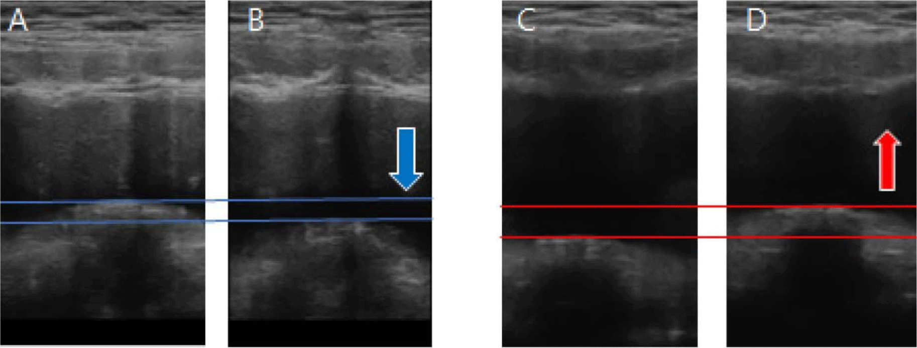
PFMC was confirmed by transabdominal ultrasound imaging using a portable ultrasonography device (SONON 300L; Healcerion, Seoul, Korea) with a curved transducer (3.5 MHz), which has been validated.20 Prior to testing, a bladder filling protocol was implemented to ensure that the subjects had sufficient fluid in their bladders to allow clear imaging, such that the measurements could be recorded. To fill the bladder, the subjects consumed 450–500 mL of water in a 1h period half an hour prior to the testing time. PFM thickness was assessed under ultrasound visualization (7.5 MHz straight linear array transducer; Dornier MedTech, Munich, Germany). For transverse plane transabdominal ultrasound imaging of the bladder base, the ultrasound transducer was placed in a transverse orientation, across the middle of the abdomen and immediately superior to the pubic symphysis. The angle of the transducer was manipulated until it was approximately 60° from vertical; it was then aimed toward the base of the bladder. The angle of the ultrasound transducer was adjusted until there was a clear image of the bladder and midline pelvic floor structures (urethra, perineal body, and rectum). The subjects were instructed to contract their pelvic floor muscle by the description “squeeze and lift your pelvic floor muscles as if trying to stop the flow of urine”. Elevation of the bladder base means the PFMC, and the subjects received feedback on PFMC through ultrasound images (Figure 2).
The Trigno wireless EMG system (Delsys Inc., Boston, MA, USA) was employed to assess the EMG activity of the TrA/IO, EO, and multifidus (MF) muscles on both sides. The EMG system was instrument with proven validity.21 The sampling rate was set at 1,000 Hz, with a bandpass filter ranging from 20 to 450 Hz. All raw EMG data underwent conversion into root mean square data for subsequent analysis. Electrodes for the TrA/IO, EO, and MF were positioned following Criswell’s guidelines.22 Prior to electrode placement, the skin was shaved and cleaned with alcohol and cotton to minimize skin impedance. To standardize the EMG activity of the TrA/IO (positioned 2 cm inferiorly and medially to the anterior superior iliac spine), EO (approximately 15 cm laterally to the umbilicus), and MF (at the L5 level aligned parallel to the line between the posterior superior iliac spine and L1-L2 interspace) muscles, the maximum voluntary isometric contraction (MVIC) of these muscles was assessed using previously suggested maneuvers.23 MVIC values were determined via manual muscle testing to normalize EMG values for the TrA/IO, EO, and MF muscles.24 Each muscle underwent two MVIC trials with a one-minute rest period between trials. The average EMG value of the middle 3 seconds of the MVIC trials was used for normalization purposes for each muscle. All EMG data during ASLR were presented as percentages of MVIC (%MVIC).
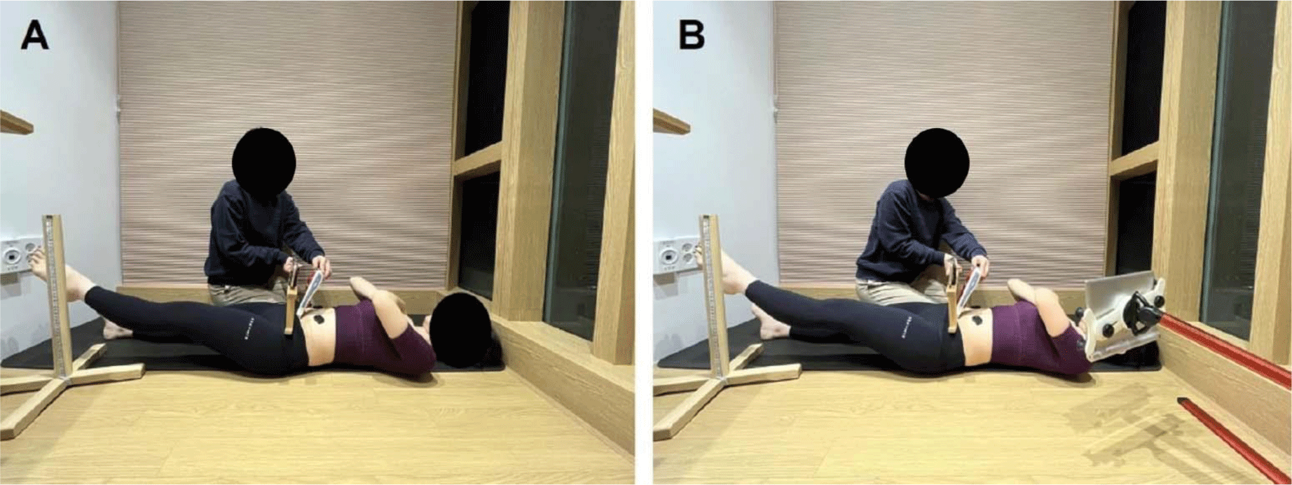
To measure pelvic transverse rotation, a smartphone was attached to the smartphone holder of the SBMT, which is a device equipped with a wooden frame designed to secure the smartphone in a position suitable for measuring the transverse rotation angle of the pelvis. Previous research has demonstrated the SBMT’s ability to provide highly reliable pelvic rotation measurements. To gauge pelvic rotation, the lower horizontal bar of the SBMT was positioned on both anterior superior iliac spines.25 An Android inclinometer application (clinometer level and slope finder; Plaincode Software Solutions, Stephanskirchen, Germany) was utilized to document the pelvic rotation angle during prone knee flexion. Before taking measurements with the SBMT, the inclinometer application underwent calibration by placing the SBMT on a flat surface. Positive angular values were characterized as rotation to the right (clockwise direction) from the subjects’ perspective.
The Active Straight Leg Raise (ASLR) was conducted with subjects lying supine and their feet positioned 20 cm apart.18 Each participant lifted their dominant leg, determined by their preference in kicking a soccer ball.26 Subjects were instructed to raise one leg until the heel was 20 cm above the table, maintaining it elevated for approximately 10 seconds (“Normal”). To enhance statistical accuracy, this procedure was repeated three times per leg. Following each ASLR, subjects were instructed to relax for about 10 seconds. The entire process was then repeated with PFMC involving ultrasound feedback (Figure 1).


The average values for the three trials of each ASLR, with and without PFMC, were utilized for all analyses. A Kolmogorov-Smirnov Z-test was conducted to assess whether continuous data followed a normal distribution. We employed a paired t-test to compare normalized EMG muscle activity between the bilateral TrA/IO, EO, and MF muscles, as well as to examine pelvic transverse rotation angle (dependent variables), with and without PFMC during ASLR (independent variables). P-values less than 0.05 were deemed statistically significant. SPSS software (ver. 16.0; SPSS Inc., Chicago, IL, USA) was employed for all data analyses. Cohen’s d was computed to evaluate the effect size between conditions with and without PFMC.
RESULTS
For this study, 16 subjects were recruited. There were 11 in nulliparae, 4 in primiparae, 1 in secundiparae. All parous subjects underwent a vaginal delivery. The dominant leg of all subjects was the right.
Activation of TrA/IO was significantly greater on both the right (p<.001) and left (p=.002) sides during ASLR with versus without PFMC. Activation of the ipsilateral EO (p=.498) was not significantly different with versus without PFMC. Activation of the contralateral EO (p=.002) was significantly lower during ASLR with compared to without PFMC. Activation of MF was not significantly different on the right (p=.099) or left (p=.139) side between ASLR with and without PFMC (Table 1).
The amount of pelvic rotation decreased significantly in ASLR with compared to without PFMC (without PFMC: 6.81±2.75 %MVIC, with PFMC: 2.49±1.65 %MVIC, p <.001) (Table 1).
DISCUSSION
PFMC was shown to be effective in improving stability of the lumbo-pelvic region. In this study, PFMC led to increased TrA/IO (23%) and decreased contralateral EO (–42%) muscle activation, together with a greater decrease in pelvic rotation angle (–57%) during ASLR. MF and ipsilateral EO muscle activation showed no significant difference with versus without PFMC during ASLR.
In this study, TrA/IO muscle activation during ASLR was greater with compared to without PFMC. The reason for this result may be co-activation of TrA/IO and PFMs. This is consistent with a previous study showing that, during PFMC, the abdominal muscle was more active in symptomatic and asymptomatic groups compared to without PFMC.10 The PFMs do not work in isolation; rather, they work in synergy with the abdominal muscles.7–9 This co-activation is necessary for the development of intra-abdominal pressure and is thought to contribute to lumbo-pelvic stability.9 Furthermore, co-activation of the IO muscle increases pelvic rim integrity, which is necessary for proper function of the suspensory system of the sacrum.27–28 Considering previously mentioned contents as a whole, PFM strengthens the control of the lumbo-pelvic. Therefore, co-activation of PFMs and TrA/IO muscles may result in optimal weight transfer through the lumbo-pelvic region during ASLR. In addition, Synergistic co-activation of the PFM and TrA/IO is likely that contributes to urinary continence.
In this study, compared to ASLR without PFMC, ASLR with PFMC decreased muscle activation of the contralateral EO. One possible explanation for this is force transmission through the anterior oblique sling, which consists of the ipsilateral IO and contralateral EO.29 Such a myofascial sling functions as a series of anatomically linked muscles, assisting in the transfer of force throughout the trunk, notably from the lower to the upper body.30–31 However, any dysfunction of the lumbo-pelvic region impairs myofascial force and energy transmission across the slings. For an ASLR, the hip flexors pull the ilium forward.32 TrA/IO muscle activity may cause the pelvis to rotate backward, thus contributing to inhibition of the anterior rotation of the ipsilateral ilium. Thus, TrA/IO co-contraction through PFMC may efficiently transfer a force to the contralateral EO by stabilizing the lumbo-pelvic region through anterior oblique sling. Consequently, the contralateral EO requires less force to stabilize the lumbo-pelvic region. This results in lower EO muscle activity compared to without PFMC.
Regarding the activities of the ipsilateral EO and both MF muscles during ASLR, there was no significant difference with versus without PFMC. The reason for this may be that the ipsilateral EO is already playing a more significant role in maintaining posture than the co-contraction resulting from the PFMC. Beales et al.33 reported that, when lifting the leg, the majority of their subjects experienced a shift from contralateral phasic activation to ipsilateral tonic activation (i.e., adopted a bracing strategy) of the EO and chest wall on the side of the ASLR. This bracing strategy aligns with the increased EO activation observed during ASLR in pregnant subjects with CPP compared to pain-free pregnant women.34 Therefore, the ipsilateral EO, which had already adopted the bracing strategy in response to the physical load of lifting the leg, was not affected by the co-contraction induced by PFMC. The MF also acts more as a tonic muscle during functional movement than the co-contraction resulting from the PFMC; based on previous studies reporting continuous activity of MF during standing and gait, the MF has a role in tonic posture.35–36 Therefore, the MF muscles may show no significant differences between before and after PFMC, because they may play a role in tonic posture rather than being co-contracted (with PFMC) during ASLR.
In this study, the amount of lumbo-pelvic rotation was significantly lower during ASLR with than without PFMC. Decreased lumbo-pelvic rotation during ASLR with PFMC may be caused by synergistic co-contraction of the core muscles. Previous studies have improved our understanding of the synergistic co-activation of the abdominal muscles and PFMs that gives rise to intra-abdominal pressure and allows for load transfer.7 Sapsford et al.8 observed concurrent abdominal muscle recruitment when a PFMC was performed in continent women. Automatic activation of the abdominal muscles was seen when the PFMs were voluntarily contracted. Accordingly, the PFMs are generally accepted as part of the trunk stability mechanism.37 According to previous studies, abdominal muscle co-contraction by PFMC may improve the internal stability of the lumbo-pelvic region, resulting in reduced pelvic transverse rotation.
This study had several limitations. Firstly, we did not assess the activity of the diaphragm and PFM as indicators of primary trunk and pelvic stabilization. Secondly, our study only included healthy women, limiting the generalizability of our results to other demographic groups including CPP. Finally, we did not consider the contact of the EMG electrode sensor of the MF with the floor. It is possible that the EMG sensor contacting the floor could affect the EMG data. Additional research is necessary to investigate the impact of ASLR with PFMC in individuals suffering from lumbo-pelvic pain. While our findings may not be extrapolated to men, existing literature indicates that men tend to exhibit greater lumbo-pelvic rotation and experience more pain during limb movement tests compared to women.
CONCLUSIONS
The activity of the TrA/IO muscles increased, while the activity of the contralateral EO and the amount of pelvic rotation decreased, during ASLR with compared to without PFMC. These results suggest that the PFMC method may be effective for decreasing unwanted pelvic rotation and increasing TrA/IO muscle activity during ASLR exercise.


