


INTRODUCTION
The primary role of the gluteus medius (Gmed) is to control the hip joint’s abduction movement and provide lateral stability of the hip joint and pelvis.1,2 Hence, strengthening exercises for the Gmed are important for functional movement of the lower limbs.3 The hip abductors must provide support to stand on one leg, and the Gmed plays a key role.4 In addition, weakness of the Gmed can cause back pain, and dysfunction of the Gmed affects various musculoskeletal disorders.5,6 Therefore, the Gmed provides balance between the body and lower limbs by controlling the movement of the hip joint.7 Strengthening the Gmed is one of the main goals of clinicians during the rehabilitation process for lower limb and back injuries.8
The clam exercise (CE) is recommended to strengthen the Gmed.9,10 The CE, frequently used in hip joint training for rehabilitating the lower back and lower extremities, combines hip joint abduction and lateral rotation movements performed in a side-lying position.10 It increases the Gmed’s activity while inhibiting the tensor fascia lata’s (TFL) activity.9 In addition, the CE is a hip joint strengthening exercise that selectively activates the gluteus maximus (Gmax).10 However, to perform the CE effectively, the patient must be able to control the movement of the lumbar pelvis himself, which is influenced by the lumbar-pelvic stability.11,12
The muscles of the abdomen control trunk movement and play an essential role in improving the stability and function of the lumbar pelvis.13,14 The abdomen muscles maintain the alignment of the lumbar pelvis.13 At the same time, the arms and legs move, suppress compensatory movements in the torso to enable efficient movement, and prevent mechanical stress from being given to the spinal joints.15 Weakness of the abdominal muscles increases lumbar lordosis and anterior tilt of the pelvis, resulting in instability of the lumbar segment and changes in the wrong lumbar-pelvic movement pattern.16,17 Therefore, strengthening the abdominal muscles is essential to maintaining normal spinal alignment and providing spinal segmental stability.4
As an exercise method to evaluate lumbar-pelvic stability, the abdominal draw-in maneuver (ADIM) improves lumbar- pelvic stability by selectively contracting the transverse abdominis (TrA) and multifidus.14,18 The ADIM improves the proactive postural control function by improving the delayed onset of muscle contraction of the TrA and multifidus and increases the lumbopelvic stability by increasing the coordination with local and global muscles.19 The ADIM also prevents anterior tilting of the pelvis and reduces lumbar lordosis.17
In a previous study, it was reported that an increase in lumbar-pelvic stability during side-lying hip abduction exercises selectively strengthened the Gmed.20–22 However, that study was conducted only for the general population, and no study on the CE for subjects with weak abdominal muscles (WAM) has been undertaken. Chan et al.11 reported that the activation of the abdominal core using the abdominal bracing maneuver increased the activity of the Gmed during the CE. However, studies on the effect of the ADIM on the CE are still lacking. Therefore, this study was conducted to determine the muscle activity of the hip joint muscles during the CE after performing the ADIM in men with WAM. We hypothesized that the TFL activity would be significantly decrease and the Gmed and Gmax activities would be significantly increase during CE after performing the ADIM in men with WAM.
METHODS
Subjects included 11 healthy men and 11 men with WAM. All participants resided in Dae-gu city (Table 1). All subjects were subjected to a trunk flexion manual muscle strength test using Kendall’s method by the principal author of this study, and subjects with WAM strength were selected who had a grade of less than poor.23 Those with dysfunction or pain in the spine, pelvis, or legs; those who regularly exercised for strength; and those with deformities were excluded from this study. The purpose of this study and the experimental method was thoroughly explained, and the experiment was conducted only for those who voluntarily consented to participate in accordance with the ethical principles of the Declaration of Helsinki. This study complied with the research ethics regulations specified by the Institutional Review Board of the Dae-gu University.
TeleMyo DTS EMG (Noraxon Inc., Scottsdale, AZ, USA), a wireless surface electromyography device, was used to measure the internal oblique (IO), Gmed, Gmax, and TFL activity during the CE. Disposable single-surface electrodes made of Ag and AgCl were used in the experiment. To minimize skin resistance, hair was removed with a thin razor and the stratum corneum was removed by rubbing with fine sandpaper 2 to 3 times. To remove oil from the skin surface, the skin was wiped with an alcohol swab 2 to 3 times, and then electrodes were attached to each muscle. The location of the electrodes for each muscle is shown in Table 2.11,24
EMG signal processing was analyzed using Myo-Research Master Edition 1.06 XP software. The sampling rate was 1,500 Hz, the frequency bandwidth was 20 to 400 Hz, and a notch filter was used to remove 60 Hz noise. All collected EMG signals were processed as root mean square (RMS). The collected signals from each muscle were normalized to the percentage of maximum voluntary isometric contraction (%MVIC). The MVIC measurement posture of the target muscles was measured using Kendall’s method.23 The MVIC for each muscle was performed for 5 seconds, and the average value was measured three times for 3 seconds, excluding the first and last second, and the average value was used.
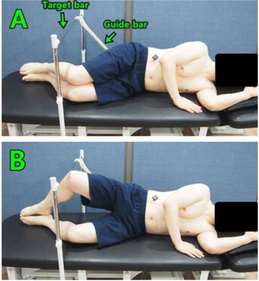
Before the intervention, the muscle activity of the IO, Gmed, Gmax, and TFL during the CE was measured for each subject. The subject was lying on his side, with the hip flexion at 45° and the knee joint flexion at 90°, with both legs touching, maintaining a straight spinal alignment, and performing the CE without both feet moving apart (Figure 1). A target bar was installed, and the hip joint abduction angle was set to 30°.11 A guide bar was installed to prevent backward rotation of the waist and pelvis, and if it touched the guide bar, it was invalidated.25 Participants were asked to maintain their legs in contact with the target bar for 5 seconds, and data collected for 3 seconds, excluding the first and last seconds, were used for analysis. All measurements were performed three times, with a 30-second rest period between measurements, and the average value was used for data analysis.
After measuring the muscle activities, the ADIM was fully explained to the subjects, and the ADIM was performed using a pressure biofeedback device (STABILIZERTM, Chattanooga Group Ontario, Canada). The ADIM was performed while supine, with both feet shoulder-width apart and with the hip flexion at 45° and the knee flexion at 90°.21 To maintain the lumbar-pelvic curve, a pressure biofeedback unit was placed on the subject’s lumbar region, the pressure was set to 40 mmHg, and the subject was instructed to pull the navel toward the lumbar spine while exhaling. At this time, the pressure gauge was set to increase by 2 to 3 mmHg. The participant held the navel pulled for 5 seconds, followed by a 5-second rest. This was repeated 10 times, and 10 repetitions were designated one set for five sets. To prevent muscle fatigue, a 20-second rest period was allowed for each set. The total exercise time was 10 minutes.
After subjects in each group performed the ADIM, the activity of the muscles was measured during the CE using the same method as before the intervention.
The data from this experiment were subjected to statistical analysis using the PASW® Statistics 18 software. The Shapiro-Wilk test was performed as a normality test to confirm normal distribution. A paired t-test was used to determine the difference between groups before and after exercise, and an independent t-test was used to compare differences between groups in the amount of change before and after exercise. The statistical significance level was set at .05.
RESULTS
The intra-group comparison results for muscle activity of the IO, Gmed, Gmax, and TFL before and after intervention are shown in Table 3. The IO, Gmed, and Gmax muscle activity significantly increased after the intervention in the WAM group (p<0.05). The TFL muscle activity significantly decreased after the intervention in the WAM group (p<0.05). In the control group, the activity of all muscles showed no significant difference after the intervention compared to before the intervention (p>0.05). (Table 3)
In the comparison between groups before the intervention, TFL activity was significantly higher in the WAM group than control group (p<0.05). The activity of the IO, Gmed, and Gmax had no significant difference (p>0.05) (Figure 2)(Table 4).
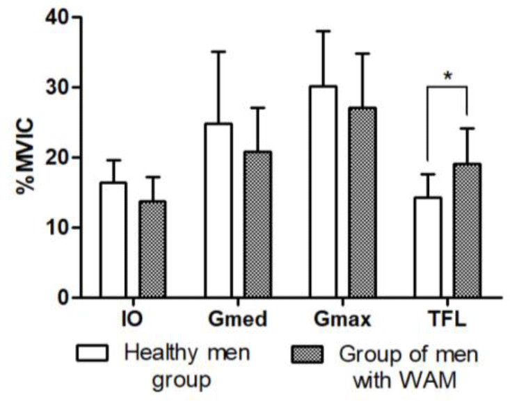
The amount of change in the activity of Gmed and TFL showed a significant difference when compared between groups (p<0.05), while the amount of change in the activity of the IO and Gmax showed no significant difference (p> 0.05)
DISCUSSION
This study was conducted to determine the effect of the IO, Gmed, Gmax, and TFL activity during the CE after ADIM for 10 minutes in healthy men and men with WAM.
The ADIM increases lumbar-pelvic stability and selectively strengthens the TrA.14,18 Weakness of the abdominal muscles causes an unstable lumbar pelvis and reduces the activity of the TrA.17,26 Using the muscle activity measurement method of the TrA performed in a previous study; TrA activity was indirectly confirmed in this study by measuring the IO activity11,27,28. In this study, IO significantly increased after the intervention within the WAM group. The reason for these results is that in men with WAM, the ADIM promoted the activity of the TrA, which may not have been used correctly. Also, it is thought that the TrA, whose activity increased through motor learning, was transferred during the CE, thereby increasing the muscle activity of the IO. In this study, there was no significant difference in the within-group comparison among healthy men, which is thought to be because healthy men had no problems with the activity of the TrA before the intervention.
In this study, TFL activity before intervention was significantly higher in the WAM group than in the control group. Neumann4 suggested that abdominal weakness could cause excessive TFL activity. For this reason, it is believed that the TFL activity was high in the WAM group before the intervention in this study. In Choi & Lee’s29 study, normal subjects and patients with lumbar-pelvic instability were compared during the CE. The group of unstable patients showed significantly lower Gmed activity, but there was no significant difference in TFL activity. While Choi & Lee’s29 study allowed compensatory pelvis rotation during the CE, this study maintained a neutral pelvis. Also, the hip joint flexion and abduction angles, and experimental participants differed between the two studies. It is believed that these factors caused the difference in the results.
In this study, in a comparison within the WAM group, TFL activity significantly decreased, and Gmed and Gmax activity significantly increased after intervention. Additionally, in the inter-group comparison of the amount of change before and after the intervention, there was a significant difference in the amount of change in TFL and Gmed activity. The reason for these results is thought to be that lumbar-pelvic stability was improved by increasing the IO activity due to the ADIM effect. As a result, it is believed to reduce the compensatory activity of the TFL and increase the selective activity of the Gmed and Gmax during the CE. Chan et al.11 reported that the CE using abdominal activation significantly increased Gmed activity, like this study’s results. Willcox and Bruden10 stated that increased pelvic stability can reduce TFL activity during the CE. In his research, he reported that maintaining a neutral pelvis during the CE optimizes the recruitment of the Gmed and Gmax. Koh et al.30 also said keeping a neutral pelvis during the CE increases the Gmax activity. Considering the results of previous studies and this study, it is thought that it is important to restore the stability of the lumbar pelvis and increase the action of abdominal muscles to reduce the compensatory action of the TFL and increase the activity of the Gmed and Gmax during CE.
Although the intervention period in this study was not long at 10 minutes, there was a significant change in the activity of all muscles after the intervention in the WAM group. Still, there was no significant difference in the control group. Macpherson & Watson31 reported that after 5 minutes of the ADIM, the thickness of the TrA significantly increased in functional postures such as forward arm stretching and standing while holding an object forward. In addition, measurements were retaken five months later, and it was reported that the thickness of the TrA continued. Chance-Larsen et al.32 reported that selective contraction training of the Gmax using the ADIM for 8 minutes and 20 seconds accelerated the onset of muscle contraction of the Gmax. Kim & Park33 reported that 10 minutes of the ADIM in women with WAM reduced the compensatory activity of the erector spinae during prone hip extension. Considering the results of previous studies and this study, it is believed that even a brief period of motor learning can modify movement strategies. It is thought that more effective motor learning can be expected if muscles that have not been recruited well are directly controlled.
This study has limitations. It was conducted only on adult men in their 20s, making it difficult to generalize to other age groups or patients. The number of subjects was small, with only 11 people in each group. The TrA was indirectly affected by the IO. The study was conducted as a cross-sectional study with a short intervention period, confirming only immediate effects. In future research, long-term evaluation of patients with back pain at various ages seems necessary.
CONCLUSIONS
This study compared the effects on muscle activity of the IO, Gmed, Gmax, and TFL during the CE after 10 minutes of the ADIM in healthy men and men with WAM. As a result of the study, in the men with WAM, the activity of the IO, Gmed, and Gmax significantly increased after intervention, and the TFL activity significantly decreased. In a comparison between groups, the amount of change in the activity of the TFL and Gmed was more significant in the WAM group than in the control group. Therefore, we suggest that performing the CE after the ADIM effectively reduces the compensatory activity of the TFL and increases the Gmed and Gmax activity in men with WAM.