INTRODUCTION
The trapezius muscle acts as an important dynamic function in creating and supporting functional activities in the shoulder complex.1 The upper trapezius (UT) acts to draw the acromion, clavicle, and scapula spine posteriorly and medially2 and to extend the neck and elevate the scapula.3 Other authors have suggested that the primary function of the lower trapezius (LT) is scapular depression, adduction, thoracic rotation, and extension.3 The LT is considered as an important muscle for the maintaining proper posture alignment and glenohumeral function. However, force imbalances of the UT and LT can occur abnormal scapular motion.4
The force imbalances result in subacromial impingement and rotator biceps tendonitis or cuff, which can alter acromioclavicular joint forces, and tend to degenerative changes.4 Cools et al.4 demonstrated that excessive UT activity combined with decreased LT activity cause abnormal scapular motion, which may result in subacromial impingement syndrome. Increased UT activity is generally accepted to contribute to subacromial impingement,4-9 but inconsistent findings with regard to LT activity have been reported in these patients. Consequently, minimizing muscle activity of UT with selective LT activity would be advantageous.
Although isolating muscle is a fundamental principle for manual muscle testing,3 little evidence is available to determine the ideal method to assess or increase the strength of the LT muscle, which isolated from the other part of the trapezius muscle. Representative exercises include the modified prone cobra, wall slide exercise, prone V-raise, and backward rocking diagonal arm lift.10-11 The prone V-raise is used by physical therapists while manual muscle testing of the LT muscle.3 Garcia et al. suggested that wall slide exercise produces the highest peak EMG for the lower trapezius than the other exercises.12 Among five different exercises, Arlotta et al.13 reported that the modified prone cobra is the most effective exercise to targeting strengthening of the LT muscle. Ha et al.11 showed that the backward rocking diagonal arm lift showed significant greater activity in the LT muscle compared to the other exercises.
Therefore, the primary aim of this study was to compare the UT and LT muscle activities while four exercises using the method described by Kendall et al.,3 and three other positions, which are the common clinical envirmonment.11-13 We hypothesized that the LT and UT muscle activities would be differ among the four specific exercises. If the most beneficial exercise are identified for activating the LT muscle, more effective rehabilitative program will be designed by clinicians.
METHODS
Fifteen healthy male subjects were recruited for this study (age: 23.6±1.9 years, height: 174.5±4.3 cm, weight: 68.5±8.9kg). The inclusion criteria were able to easily perform full flexion, full abduction, and full scaption; and levator scapulae, pectoralis minor, and rhomboid muscles within the normal length determined using the muscle length test.14-15 The exclusion criteria were (1) shoulder surgery or current shoulder pain (2) history of musculoskeletal, neurological, or cardiopulmonary disease. Prior to the experiment, the investigator explained the procedure to the subjects, and all subjects provided written informed consent. This study was approved by the institutional review board of Inje University (No. 2022-04-044-001). The estimation of sample size was calculated with G*Power 3.1.9.7 for Windows (University of Dusseldorf, Dusseldorf, Germany). Power analysis determined that at least 15 sub-jects were required to obtain a power of 0.8, and signifi-cance level of .05.16
Before electromyography (EMG), we performed a test to determine the dominance of the arms; the dominant arm was defined as the throwing side during sports performance.17 A Trigno wireless EMG system (Delsys, Inc., Boston, MA, USA) was used to assess the muscle activity of the dominant UT and LT. The EMG signals were sampled at 1,000 Hz with a 20-450 Hz bandpass filter. The raw EMG data were converted to root mean square data. The electrodes of the UT and LT muscles were placed in accordance with Criswell.18 The each electrode site was shaved and cleaned to decrease skin impedance. For normalization, the maximum voluntary isometric contraction (MVIC) of the muscles was measured using method presented previously.19 The MVIC trials were carried out three times for 5 s, and middle 3 s of three trials was used to obtain steady-state results. All EMG data during four specific therapeutic exercises are expressed as percentages of the MVIC (%MVIC).
After attaching the electrodes, all participants performed the modified prone cobra, wall slide exercise, prone V-raise, and backward rocking diagonal arm lift. Subjects were asked performing three trials of four different exercises considered to effectively activate the fibers of the LT muscle. Before testing, several practice trials were performed to familiarization with the exercises. The exclusion criteria were those who could not do requested exercise, but all participants carried out all the exercises correctly. All exercises remained isometric contraction for 5 s while data were recorded, with a minimum of 30 s of rest between contractions. The order of the exercises was randomized among participants. The four exercises were as follows:
-
Modified prone cobra12: The participant was lying face down on the floor with his arm side and his fingers pointing toward his toes. The individual performed trunk extension, to raise his chest approximately 10 cm off the floor. Then, the participant pulled shoulder blades back and down with thumb facing the ceiling as if the fingertips were toward the feet (Figure 1).
-
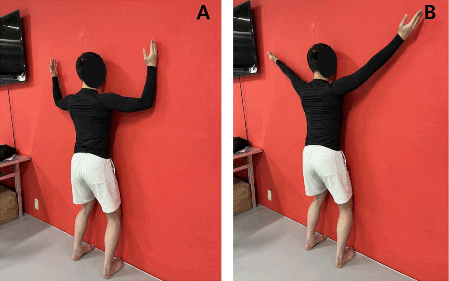
Wall slide exercise11,15,20: The participant stood facing a wall with the wall from nose to knees with feet shoulder-width apart. Then, the ulnar border of the forearms contacted the wall with shoulder 90° abduction and elbow 90° flexion. The subject was sliding his arms against the wall. The sliding motion was ended until the shoulder 145° abduction. Then, the subject lifted both hands with the elbows extended and remained the arm position (Figure 2).
-
Prone V-raise3,21: The participant was lying face down on the floor with shoulder 180° flexion at a shoulder 120° abduction and with elbows extended. The participant lifted off his arms from the floor to ear level with thumbs pointing toward the ceiling (Figure 3).
-
Backward rocking diagonal arm lift11: The subject was instructed to performing the quadruped position and then rocking backward. The investigator abducted the subject’s shoulder to 145° and instructed the subject lifting the dominant arm with the elbows extended up to his shoulders flexed to 180° (Figure 4).

The EMG activation recorded from the LT muscle relative to that recorded from both the LT and UT muscles was computed for each trial using the following equation:
Perfect LT/UT ratio of the lower trapezius muscle (LT/UT ratio = 100%) would result when there was no activity of the upper fibers of the trapezius muscle during an exercise.
A one sample Kolmogorov-Smirnov test was used to investigate the normality of the distribution of the EMG. A one-way repeated-measures analysis of variance (ANOVA) was used to test for differences in the UT and LT muscle activities and LT/UT ratios among the four exercises. Significant main effects were followed up using the Bonferroni post hoc test. All statistical analyses were conducted with SPSS Statistics version 18 (SPSS Inc., Chicago, IL, USA), and α<.05 was taken to indicate statistical significance. If a significant interaction was found, the Bonferroni correction was set at .017 (.05/3) at a statistically significant level.
RESULTS
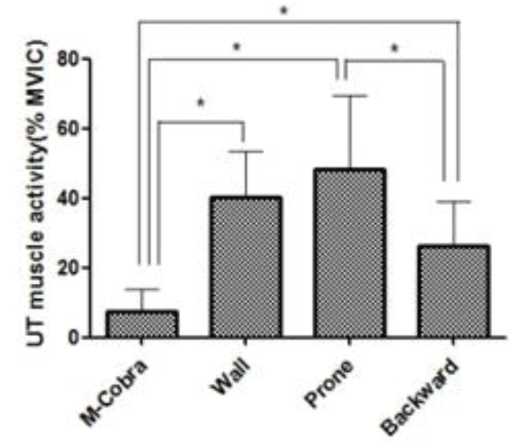
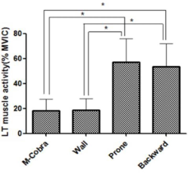
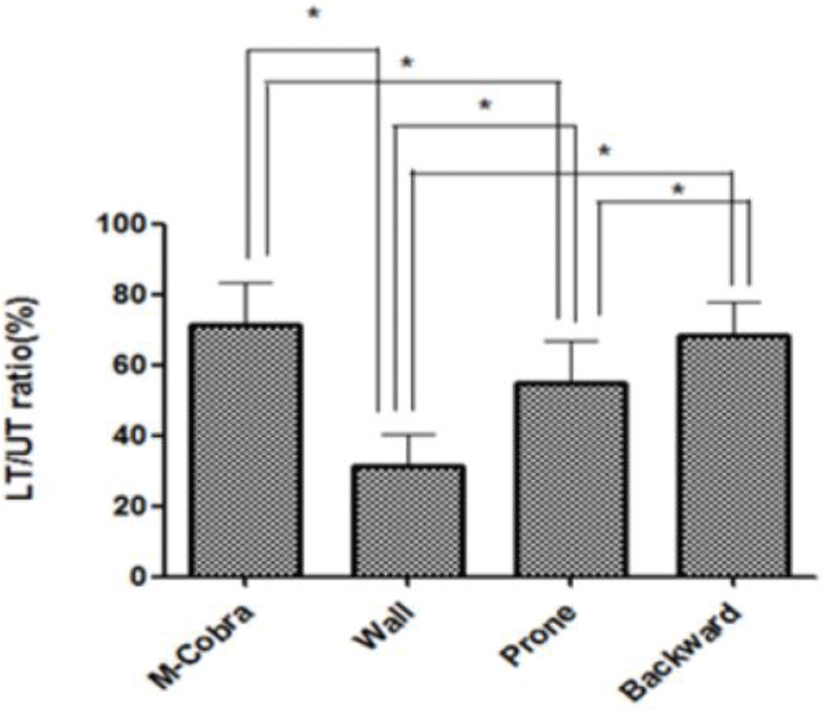
In this study, 15 healthy male subjects were recruited without missing data. The dominant arm of all subjects was the right. The results of statistical analyses are shown in Table 1. UT muscle activity was significantly lower during the modified prone cobra than during the other exercises (p<.001), followed by the backward rocking diagonal arm lift (Figure 5). LT muscle activity was significantly greater during the backward rocking diagonal arm lift than the wall slide exercise (p<.001) or modified prone cobra (p<.001), and was not significantly different from that during the prone V-raise (p=1.000) (Figure 6). The LT/UT ratio was significantly greater during the backward rocking diagonal arm lift than during the wall slide exercise (p<.001) or prone V-raise (p<.001), and was not significantly different from that during the modified prone cobra (p=1.000) (Figure 7).


DISCUSSION
We investigated UT and LT muscle activity during four specific therapeutic exercises. LT muscle activity was sig-nificantly greater during the backward rocking diagonal arm lift than during the wall slide exercise or modified prone cobra, and was not significantly different from that during the prone V-raise. The LT/UT ratio was significantly greater during the backward rocking diagonal arm lift than during the wall slide exercise or prone V-raise, and was not significantly different from that during the modified prone cobra. Finally, UT muscle activity was significantly lower during the modified prone cobra than during the other exercises, followed by the backward rocking diagonal arm lift.
LT muscle activity was significantly greater during the backward rocking diagonal arm lift than during the wall slide exercise (183.62% improvement) or modified prone cobra (192.61% improvement), and was not significantly different from that during the prone V-raise, which caused the greatest activation. These results are consistent with previous findings. Ha et al.11 and Yong & Weon22 reported that LT muscle activity was significantly greater during the backward rocking diagonal arm lift than that during the wall slide exercise and modified prone cobra, respectively. These findings can be explained as the backward rocking diagonal arm lift is performed in an antigravity position and “diagonal overhead” arm position in line with the LT. However, LT muscle activity during the backward rocking diagonal arm lift was not significantly different from that during the prone V-raise in the present study, which is inconsistent with the results of Ha et al.11 who reported that the backward rocking diagonal arm lift showed significantly more activity in the LT muscle than the prone V-raise. This difference was likely because several subjects in this study did not overcome the scapular anterior tilt. For the backward rocking diagonal arm lift, the flexed thoracic position results in a scapular anterior tilt, which make a difficult the scapular posterior tilt. The LT muscle is highly activated when overcoming a challenging posture, otherwise it becomes less active.23 Therefore, if the subject did not overcome the scapular anterior tilt, LT muscle activity may not have been significantly different from that in the prone V-raise or may have been rather low. Based on previous studies, the backward rocking diagonal arm lift is an beneficial method for activation of the LT muscle because it challenges the scapular anterior tilt, and it will be a more effective method with monitoring of scapular posterior tilt.
The LT/UT ratio was significantly greater during the backward rocking diagonal arm lift than during the wall slide exercise (117.77% improvement) or prone V-raise (24.65% improvement), and was not significantly different during the modified prone cobra, which had the greatest activation. One possible explanation is that the backward rocking diagonal arm lift position involves slight cervical flexion compared to the other exercise positions. De Lorenzo et al.24 suggested immobilization in a position of slight neck flexion, implying that the sarcomeres of the cervical mul-tifidus are of optimal length in a slightly flexed posture.25 Previous studies suggested that a neutral cervical posture reduced altered activation of the UT muscles.26-28 Therefore, UT muscle activity was lower during the backward rocking diagonal arm lift than the other exercises due to the neck posture, which may have increased the LT/UT ratio. However, there was no significant difference in the LT/UT ratio between the backward diagonal arm lift and modified prone cobra, which had the greatest activation. The modified prone cobra posture causes slight depression and adduction of the scapula without upward rotation. In this posture, the muscle length is long for the UT and short for the LT, which increases the LT/UT muscle activity ratio. However, the LT muscle activity during the modified prone cobra was 18.28± 9.30%MVIC, which is insufficient to strengthen the muscle. In most rehabilitation protocols, exercises that produce more than 40%–60% of the MVIC are desired during advanced phases of shoulder rehabilitation.29 Furthermore, the modified prone cobra position did not include functional move-ment of the LT (scapular upward rotation). Activation of a specific muscle is increased during intended functional action of the muscle, which is due to stimulation of the muscle fibers.30-31 Taken together, these observations suggest that the backward rocking diagonal arm lift may be a more effective exercise than the modified prone cobra for decreasing UT muscle activity and increasing LT muscle activity.
Finally, UT muscle activity was significantly lower during the modified prone cobra than during the other exercises, followed by the backward rocking diagonal arm lift. This is consistent with a previous report showing that the modified prone cobra produced significantly less isolation of the UT than the prone row exercise.13 However, the modified prone cobra is performed below 90° of humeral elevation. Moseley et al.32 reported that upper arm moving in an arc between 90° and 150° elevation most effective for recruiting LT during glenohumeral abduction and rowing exercises. The muscle fibers of the LT have been estimated to run at an angle of approximately 125°.21 They also reported that the LT showed superior activation with the arm in line with the muscle fibers of the LT. Based on previous studies, the backward rocking diagonal arm lift may be an beneficial method to activating the LT while decreasing UT activation as the exercise was performed at 145° of humeral elevation in line with the LT and showed the second smallest level of UT activation after the modified prone cobra. In addition, UT muscle activity during the backward rocking diagonal arm lift was 26.13±12.83 (%MVIC) at relatively weak contractions (10–30%MIVIC).
This study had several limitations. First, we did not confirm the scapular posterior tilt during each exercise. Additional studies are necessary to determine the correlation between the amount of scapular posterior tilt and other exercises. Second, all subjects were asymptomatic young men. Future studies including symptomatic and/or older subjects are required to generalize our findings. Finally, the cross-talk over the trapezius muscles has not been reported. In the trapezius muscle, cross-talk can potentially occur from deeper muscles such as the supraspinatus, or the rhomboid muscle.
CONCLUSIONS
The present study measured UT and LT muscle activity during four specific therapeutic exercises. In terms LT muscle activity and the LT/UT ratio, the backward rocking diagonal arm lift was not significantly different from the other exercises, which produced greater levels of activation. In terms of UT muscle activity, the backward rocking diagonal arm lift produced the second smallest degree of activation after the modified prone cobra, which produced the lowest level of activation. The backward rocking diagonal arm exercise should be considered an effective strengthening exercise for the LT muscle, while decreasing UT muscle activity. The scapular posterior tilt should be confirmed during exercise testing.


